 Movie
Movie Controller

[English] 日本語
 Yorodumi
- PDB-4etj: Crystal Structure of E6H variant of de novo designed serine hydro... -
+ Open data

- Basic information
| Entry | Database: PDB / ID: 4etj | ||||||
|---|---|---|---|---|---|---|---|
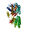
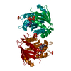
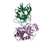
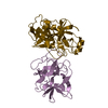
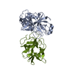
| Title | Crystal Structure of E6H variant of de novo designed serine hydrolase OSH55, Northeast Structural Genomics Consortium (NESG) Target OR185 | ||||||
 Components Components | Serine hydrolase | ||||||
 Keywords Keywords | DE NOVO PROTEIN / Structural Genomics / PSI-Biology / Protein Structure Initiative / Northeast Structural Genomics Consortium / NESG | ||||||
| Function / homology | Leucine Aminopeptidase, subunit E, domain 1 / Leucine Aminopeptidase, subunit E; domain 1 / 3-Layer(aba) Sandwich / Alpha Beta / PHOSPHORYL-HEXAETHYLENE GLYCOL Function and homology information Function and homology information | ||||||
| Biological species | artificial gene (others) | ||||||
| Method |  X-RAY DIFFRACTION / SYNCHROTRON / MIR / Resolution: 2.203 Å X-RAY DIFFRACTION / SYNCHROTRON / MIR / Resolution: 2.203 Å | ||||||
 Authors Authors | Kuzin, A. / Su, M. / Seetharaman, J. / Kornhaber, K. / Kornhaber, G. / Rajagopalan, S. / Baker, D. / Everett, J.K. / Acton, T.B. / Montelione, G.T. ...Kuzin, A. / Su, M. / Seetharaman, J. / Kornhaber, K. / Kornhaber, G. / Rajagopalan, S. / Baker, D. / Everett, J.K. / Acton, T.B. / Montelione, G.T. / Tong, L. / Hunt, J.F. / Northeast Structural Genomics Consortium (NESG) | ||||||
 Citation Citation | Journal: Nat.Chem.Biol. / Year: 2014 Title: Design of activated serine-containing catalytic triads with atomic-level accuracy. Authors: Rajagopalan, S. / Wang, C. / Yu, K. / Kuzin, A.P. / Richter, F. / Lew, S. / Miklos, A.E. / Matthews, M.L. / Seetharaman, J. / Su, M. / Hunt, J.F. / Cravatt, B.F. / Baker, D. | ||||||
| History |
|
- Structure visualization
| Structure viewer | Molecule: MolmilJmol/JSmol |
|---|
- Downloads & links
-Download
| PDBx/mmCIF format | 4etj.cif.gz | 81.9 KB | Display | PDBx/mmCIF format |
|---|---|---|---|---|
| PDB format | pdb4etj.ent.gz | 59.2 KB | Display | PDB format |
| PDBx/mmJSON format | 4etj.json.gz | Tree view | PDBx/mmJSON format | |
| Others |  Other downloads Other downloads |
-Validation report
| Arichive directory | https://data.pdbj.org/pub/pdb/validation_reports/et/4etjftp://data.pdbj.org/pub/pdb/validation_reports/et/4etj | HTTPS FTP |
|---|
-Related structure data
| Related structure data |  3tp4C  3v45SC  4drtC  4essC  4etkC  4f2vC  4jcaC  4jllC  4jvvC S: Starting model for refinement C: citing same article ( |
|---|---|
| Similar structure data | |
| Other databases |
-Links
 PDBj
PDBj
- Assembly
| Deposited unit | 
| ||||||||
|---|---|---|---|---|---|---|---|---|---|
| 1 | 
| ||||||||
| Unit cell |
| ||||||||
| Components on special symmetry positions |
| ||||||||
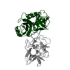
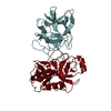
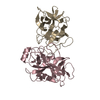
| Details | monomer,17.05 kD,95.7%, dimer,34.77 kD,4.68% |
-Components
-Protein , 1 types, 1 molecules A
| #1: Protein | Mass: 17684.277 Da / Num. of mol.: 1 / Mutation: E6H Source method: isolated from a genetically manipulated source Source: (gene. exp.) artificial gene (others) |
|---|
-Non-polymers , 5 types, 41 molecules 


| #2: Chemical | ChemComp-CL /  Mass: 35.453 Da / Num. of mol.: 1 / Source method: obtained synthetically / Formula: Cl Mass: 35.453 Da / Num. of mol.: 1 / Source method: obtained synthetically / Formula: Cl |
|---|---|
| #3: Chemical | ChemComp-PE4 /  Mass: 354.436 Da / Num. of mol.: 1 / Source method: obtained synthetically / Formula: C16H34O8 / Comment: precipitant*YM Mass: 354.436 Da / Num. of mol.: 1 / Source method: obtained synthetically / Formula: C16H34O8 / Comment: precipitant*YM |
| #4: Chemical | ChemComp-PE5 /  Mass: 398.489 Da / Num. of mol.: 1 / Source method: obtained synthetically / Formula: C18H38O9 / Comment: precipitant*YM Mass: 398.489 Da / Num. of mol.: 1 / Source method: obtained synthetically / Formula: C18H38O9 / Comment: precipitant*YM |
| #5: Chemical | ChemComp-PE6 /  Mass: 362.311 Da / Num. of mol.: 1 / Source method: obtained synthetically / Formula: C12H27O10P Mass: 362.311 Da / Num. of mol.: 1 / Source method: obtained synthetically / Formula: C12H27O10P |
| #6: Water | ChemComp-HOH / Mass: 18.015 Da / Num. of mol.: 37 / Source method: isolated from a natural source / Formula: H2O |
-Experimental details
-Experiment
| Experiment | Method: X-RAY DIFFRACTION / Number of used crystals: 1 |
|---|
- Sample preparation
| Crystal | Density Matthews: 2.74 Å3/Da / Density % sol: 55.15 % |
|---|---|
| Crystal grow | Temperature: 293 K / pH: 7 Details: Protein solution: 100mM NaCl, 5mM DTT, 0.02% NaN3, 10mM Tris-HCl (pH 7.5), Reservoir solution: 0.1M MgCl2, 0.1M Bis-Tris, 80% PEG400, microbatch under oil, temperature 293KK |
-Data collection
| Diffraction | Mean temperature: 100 K |
|---|---|
| Diffraction source | Source: SYNCHROTRON / Site: NSLS  / Beamline: X4C / Wavelength: 0.97925 Å / Beamline: X4C / Wavelength: 0.97925 Å |
| Detector | Type: MAR CCD 165 mm / Detector: CCD / Date: Mar 21, 2012 / Details: MIRROR |
| Radiation | Monochromator: Si 111 CHANNEL / Protocol: SINGLE WAVELENGTH / Monochromatic (M) / Laue (L): M / Scattering type: x-ray |
| Radiation wavelength | Wavelength: 0.97925 Å / Relative weight: 1 |
| Reflection | Resolution: 2.2→50 Å / Num. obs: 19058 / % possible obs: 100 % / Observed criterion σ(I): -3 / Redundancy: 14.9 % / Biso Wilson estimate: 36.45 Å2 / Rmerge(I) obs: 0.09 / Net I/σ(I): 57.6 |
- Processing
| Software |
| ||||||||||||||||||||||||||||||||||||||||
|---|---|---|---|---|---|---|---|---|---|---|---|---|---|---|---|---|---|---|---|---|---|---|---|---|---|---|---|---|---|---|---|---|---|---|---|---|---|---|---|---|---|
| Refinement | Method to determine structure: MIR Starting model: 3V45 Resolution: 2.203→36.958 Å / Occupancy max: 1 / Occupancy min: 1 / SU ML: 0.21 / σ(F): 1.34 / Phase error: 18.79 / Stereochemistry target values: ML
| ||||||||||||||||||||||||||||||||||||||||
| Solvent computation | Shrinkage radii: 0.73 Å / VDW probe radii: 1 Å / Solvent model: FLAT BULK SOLVENT MODEL / Bsol: 49.029 Å2 / ksol: 0.375 e/Å3 | ||||||||||||||||||||||||||||||||||||||||
| Displacement parameters |
| ||||||||||||||||||||||||||||||||||||||||
| Refinement step | Cycle: LAST / Resolution: 2.203→36.958 Å
| ||||||||||||||||||||||||||||||||||||||||
| Refine LS restraints |
| ||||||||||||||||||||||||||||||||||||||||
| LS refinement shell |
| ||||||||||||||||||||||||||||||||||||||||
| Refinement TLS params. | Method: refined / Origin x: 5.6904 Å / Origin y: 13.5459 Å / Origin z: 14.1463 Å
| ||||||||||||||||||||||||||||||||||||||||
| Refinement TLS group | Selection details: all |
