 Movie
Movie Controller

[English] 日本語
 Yorodumi
- PDB-4jca: Crystal Structure of the apo form of the evolved variant of the c... -
+ Open data

- Basic information
| Entry | Database: PDB / ID: 4jca | ||||||
|---|---|---|---|---|---|---|---|
| Title | Crystal Structure of the apo form of the evolved variant of the computationally designed serine hydrolase, OSH55.4_H1. Northeast Structural Genomics Consortium (NESG) Target OR273 | ||||||
 Components Components | serine hydrolase | ||||||
 Keywords Keywords | HYDROLASE / Structural Genomics / PSI-Biology / Protein Structure Initiative / Northeast Structural Genomics Consortium (NESG) | ||||||
| Function / homology | Leucine Aminopeptidase, subunit E, domain 1 / Leucine Aminopeptidase, subunit E; domain 1 / 3-Layer(aba) Sandwich / Alpha Beta / CITRIC ACID / RUBIDIUM ION Function and homology information Function and homology information | ||||||
| Biological species | artificial gene (others) | ||||||
| Method |  X-RAY DIFFRACTION / SYNCHROTRON / MOLECULAR REPLACEMENT / Resolution: 2.411 Å X-RAY DIFFRACTION / SYNCHROTRON / MOLECULAR REPLACEMENT / Resolution: 2.411 Å | ||||||
 Authors Authors | Kuzin, A.P. / Lew, S. / Rajagopalan, S. / Seetharaman, J. / Tong, S. / Everett, J.K. / Acton, T.B. / Baker, D. / Montelione, G.T. / Tong, L. ...Kuzin, A.P. / Lew, S. / Rajagopalan, S. / Seetharaman, J. / Tong, S. / Everett, J.K. / Acton, T.B. / Baker, D. / Montelione, G.T. / Tong, L. / Hunt, J.F. / Northeast Structural Genomics Consortium (NESG) | ||||||
 Citation Citation | Journal: Nat.Chem.Biol. / Year: 2014 Title: Design of activated serine-containing catalytic triads with atomic-level accuracy. Authors: Rajagopalan, S. / Wang, C. / Yu, K. / Kuzin, A.P. / Richter, F. / Lew, S. / Miklos, A.E. / Matthews, M.L. / Seetharaman, J. / Su, M. / Hunt, J.F. / Cravatt, B.F. / Baker, D. | ||||||
| History |
|
- Structure visualization
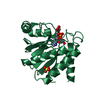
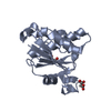
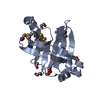
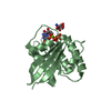
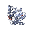
| Structure viewer | Molecule: MolmilJmol/JSmol |
|---|
- Downloads & links
-Download
| PDBx/mmCIF format | 4jca.cif.gz | 76.3 KB | Display | PDBx/mmCIF format |
|---|---|---|---|---|
| PDB format | pdb4jca.ent.gz | 56 KB | Display | PDB format |
| PDBx/mmJSON format | 4jca.json.gz | Tree view | PDBx/mmJSON format | |
| Others |  Other downloads Other downloads |
-Validation report
| Arichive directory | https://data.pdbj.org/pub/pdb/validation_reports/jc/4jcaftp://data.pdbj.org/pub/pdb/validation_reports/jc/4jca | HTTPS FTP |
|---|
-Related structure data
| Related structure data |  3tp4C  3v45C  4drtC  4essC  4etjSC  4etkC  4f2vC  4jllC  4jvvC S: Starting model for refinement C: citing same article ( |
|---|---|
| Similar structure data | |
| Other databases |


-Links
 PDBj
PDBj
- Assembly
| Deposited unit | 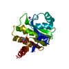
| ||||||||
|---|---|---|---|---|---|---|---|---|---|
| 1 |
| ||||||||
| 2 | 
| ||||||||
| Unit cell |
|
-Components
| #1: Protein | Mass: 17808.229 Da / Num. of mol.: 1 Source method: isolated from a genetically manipulated source Source: (gene. exp.) artificial gene (others) |
|---|---|
| #2: Chemical | ChemComp-RB /   Mass: 85.468 Da / Num. of mol.: 1 / Source method: obtained synthetically / Formula: Rb Mass: 85.468 Da / Num. of mol.: 1 / Source method: obtained synthetically / Formula: Rb |
| #3: Chemical | ChemComp-CIT /   Mass: 192.124 Da / Num. of mol.: 1 / Source method: obtained synthetically / Formula: C6H8O7 Mass: 192.124 Da / Num. of mol.: 1 / Source method: obtained synthetically / Formula: C6H8O7 |
| #4: Water | ChemComp-HOH /  Mass: 18.015 Da / Num. of mol.: 30 / Source method: isolated from a natural source / Formula: H2O Mass: 18.015 Da / Num. of mol.: 30 / Source method: isolated from a natural source / Formula: H2O |
-Experimental details
-Experiment
| Experiment | Method: X-RAY DIFFRACTION / Number of used crystals: 1 |
|---|
- Sample preparation
| Crystal | Density Matthews: 2.27 Å3/Da / Density % sol: 45.74 % |
|---|---|
| Crystal grow | Temperature: 277 K / Method: microbatch under oil / pH: 4.2 Details: Protein solution: 100mM NaCl, 5mM DTT, 0.02% NaN3, 10mM Tris-HCl (pH 7.5). Reservoir solution: RbCl 0.1M, Sodium Citrate 0.1M, PEG 1000 40%, microbatch under oil, temperature 277K |
-Data collection
| Diffraction | Mean temperature: 100 K |
|---|---|
| Diffraction source | Source: SYNCHROTRON / Site: NSLS  / Beamline: X4A / Wavelength: 0.979 Å / Beamline: X4A / Wavelength: 0.979 Å |
| Detector | Type: ADSC QUANTUM 4r / Detector: CCD / Date: Oct 2, 2012 |
| Radiation | Monochromator: Si 111 CHANNEL / Protocol: SINGLE WAVELENGTH / Monochromatic (M) / Laue (L): M / Scattering type: x-ray |
| Radiation wavelength | Wavelength: 0.979 Å / Relative weight: 1 |
| Reflection | Resolution: 2.4→30 Å / Num. obs: 11749 / % possible obs: 97.9 % / Observed criterion σ(I): -3 / Redundancy: 2.8 % / Biso Wilson estimate: 40.78 Å2 / Rmerge(I) obs: 0.06 / Net I/σ(I): 22.8 |
- Processing
| Software |
| ||||||||||||||||||||||||||||||||||||||||
|---|---|---|---|---|---|---|---|---|---|---|---|---|---|---|---|---|---|---|---|---|---|---|---|---|---|---|---|---|---|---|---|---|---|---|---|---|---|---|---|---|---|
| Refinement | Method to determine structure: MOLECULAR REPLACEMENT Starting model: 4ETJ Resolution: 2.411→29.396 Å / Occupancy max: 1 / Occupancy min: 1 / SU ML: 0.35 / σ(F): 1.36 / Phase error: 26.9 / Stereochemistry target values: ML
| ||||||||||||||||||||||||||||||||||||||||
| Solvent computation | Shrinkage radii: 0.9 Å / VDW probe radii: 1.11 Å / Solvent model: FLAT BULK SOLVENT MODEL | ||||||||||||||||||||||||||||||||||||||||
| Displacement parameters | Biso mean: 41.62 Å2 | ||||||||||||||||||||||||||||||||||||||||
| Refinement step | Cycle: LAST / Resolution: 2.411→29.396 Å
| ||||||||||||||||||||||||||||||||||||||||
| Refine LS restraints |
| ||||||||||||||||||||||||||||||||||||||||
| LS refinement shell |
| ||||||||||||||||||||||||||||||||||||||||
| Refinement TLS params. | Method: refined / Origin x: -9.9622 Å / Origin y: 1.3494 Å / Origin z: -18.8879 Å
| ||||||||||||||||||||||||||||||||||||||||
| Refinement TLS group | Selection details: all |
