 Movie
Movie Controller

[English] 日本語
 Yorodumi
- PDB-1k4s: HUMAN DNA TOPOISOMERASE I IN COVALENT COMPLEX WITH A 22 BASE PAIR... -
+ Open data

- Basic information
| Entry | Database: PDB / ID: 1k4s | ||||||
|---|---|---|---|---|---|---|---|
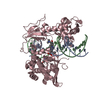
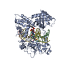
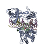
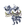
| Title | HUMAN DNA TOPOISOMERASE I IN COVALENT COMPLEX WITH A 22 BASE PAIR DNA DUPLEX | ||||||
 Components Components |
| ||||||
 Keywords Keywords | ISOMERASE/DNA / Complex (ISOMERASE-DNA) / DNA / Topoisomerase I / ISOMERASE-DNA complex | ||||||
| Function / homology |  Function and homology information Function and homology informationDNA topoisomerase / DNA topoisomerase type I (single strand cut, ATP-independent) activity / dense fibrillar component / cellular response to luteinizing hormone stimulus / embryonic cleavage / programmed cell death / supercoiled DNA binding / DNA binding, bending / response to temperature stimulus / DNA topological change ...DNA topoisomerase / DNA topoisomerase type I (single strand cut, ATP-independent) activity / dense fibrillar component / cellular response to luteinizing hormone stimulus / embryonic cleavage / programmed cell death / supercoiled DNA binding / DNA binding, bending / response to temperature stimulus / DNA topological change / rRNA transcription / SUMOylation of DNA replication proteins / animal organ regeneration / response to cAMP / response to gamma radiation / male germ cell nucleus / chromosome segregation / circadian regulation of gene expression / P-body / peptidyl-serine phosphorylation / circadian rhythm / protein-DNA complex / chromatin DNA binding / fibrillar center / single-stranded DNA binding / chromosome / double-stranded DNA binding / perikaryon / DNA replication / RNA polymerase II cis-regulatory region sequence-specific DNA binding / chromatin remodeling / response to xenobiotic stimulus / protein domain specific binding / protein serine/threonine kinase activity / chromatin binding / protein-containing complex binding / nucleolus / DNA binding / RNA binding / nucleoplasm / ATP binding / nucleus Similarity search - Function | ||||||
| Biological species |  Homo sapiens (human) Homo sapiens (human) | ||||||
| Method |  X-RAY DIFFRACTION / SYNCHROTRON / MOLECULAR REPLACEMENT / Resolution: 3.2 Å X-RAY DIFFRACTION / SYNCHROTRON / MOLECULAR REPLACEMENT / Resolution: 3.2 Å | ||||||
 Authors Authors | Staker, B.L. / Hjerrild, K. / Feese, M.D. / Behnke, C.A. / Burgin Jr., A.B. / Stewart, L.J. | ||||||
 Citation Citation | Journal: Proc.Natl.Acad.Sci.USA / Year: 2002 Title: The mechanism of topoisomerase I poisoning by a camptothecin analog Authors: Staker, B.L. / Hjerrild, K. / Feese, M.D. / Behnke, C.A. / Burgin Jr., A.B. / Stewart, L.J. | ||||||
| History |
|
- Structure visualization
| Structure viewer | Molecule: MolmilJmol/JSmol |
|---|
- Downloads & links
-Download
| PDBx/mmCIF format | 1k4s.cif.gz | 141.5 KB | Display | PDBx/mmCIF format |
|---|---|---|---|---|
| PDB format | pdb1k4s.ent.gz | 105.1 KB | Display | PDB format |
| PDBx/mmJSON format | 1k4s.json.gz | Tree view | PDBx/mmJSON format | |
| Others |  Other downloads Other downloads |
-Validation report
| Arichive directory | https://data.pdbj.org/pub/pdb/validation_reports/k4/1k4sftp://data.pdbj.org/pub/pdb/validation_reports/k4/1k4s | HTTPS FTP |
|---|
-Related structure data
| Related structure data |  1k4tC  1a36S S: Starting model for refinement C: citing same article ( |
|---|---|
| Similar structure data |


-Links
 PDBj
PDBj


- Assembly
| Deposited unit | 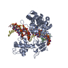
| ||||||||
|---|---|---|---|---|---|---|---|---|---|
| 1 |
| ||||||||
| Unit cell |
|
-Components
| #1: DNA chain | Mass: 3284.796 Da / Num. of mol.: 1 / Source method: obtained synthetically |
|---|---|
| #2: DNA chain | Mass: 4138.985 Da / Num. of mol.: 1 / Source method: obtained synthetically |
| #3: DNA chain | Mass: 7721.218 Da / Num. of mol.: 1 / Source method: obtained synthetically |
| #4: Protein | Mass: 70248.023 Da / Num. of mol.: 1 Fragment: Core Domain and C-Terminal Domain, Residues 174-765 Source method: isolated from a genetically manipulated source Source: (gene. exp.) Homo sapiens (human) / Production host:   Spodoptera frugiperda (fall armyworm) / References: UniProt: P11387, EC: 5.99.1.2 Spodoptera frugiperda (fall armyworm) / References: UniProt: P11387, EC: 5.99.1.2 |
| Has protein modification | Y |
-Experimental details
-Experiment
| Experiment | Method: X-RAY DIFFRACTION / Number of used crystals: 1 |
|---|
- Sample preparation
| Crystal | Density Matthews: 3.43 Å3/Da / Density % sol: 64.17 % | ||||||||||||||||||||||||||||||||||||||||||||||||||||||||
|---|---|---|---|---|---|---|---|---|---|---|---|---|---|---|---|---|---|---|---|---|---|---|---|---|---|---|---|---|---|---|---|---|---|---|---|---|---|---|---|---|---|---|---|---|---|---|---|---|---|---|---|---|---|---|---|---|---|
| Crystal grow | Temperature: 289 K / Method: vapor diffusion, sitting drop / pH: 8 Details: PEG 8000, Tris-HCl, Na/K Phosphate, KCl, DTT, VAPOR DIFFUSION, SITTING DROP, temperature 289K | ||||||||||||||||||||||||||||||||||||||||||||||||||||||||
| Components of the solutions |
| ||||||||||||||||||||||||||||||||||||||||||||||||||||||||
| Crystal grow | *PLUS Temperature: 16 ℃ / pH: 8 | ||||||||||||||||||||||||||||||||||||||||||||||||||||||||
| Components of the solutions | *PLUS
|
-Data collection
| Diffraction | Mean temperature: 100 K |
|---|---|
| Diffraction source | Source: SYNCHROTRON / Site: NSLS  / Beamline: X25 / Beamline: X25 |
| Detector | Detector: CCD |
| Radiation | Protocol: SINGLE WAVELENGTH / Monochromatic (M) / Laue (L): M / Scattering type: x-ray |
| Radiation wavelength | Relative weight: 1 |
| Reflection | Resolution: 3.2→50 Å / Num. all: 18471 / Num. obs: 18471 / % possible obs: 85.9 % / Rsym value: 0.13 |
| Reflection shell | Resolution: 3.2→3.31 Å / Mean I/σ(I) obs: 2.95 / Rsym value: 0.13 / % possible all: 69.5 |
| Reflection | *PLUS Lowest resolution: 50 Å / Num. obs: 17874 / Rmerge(I) obs: 0.13 |
- Processing
| Software |
| |||||||||||||||||||||||||
|---|---|---|---|---|---|---|---|---|---|---|---|---|---|---|---|---|---|---|---|---|---|---|---|---|---|---|
| Refinement | Method to determine structure: MOLECULAR REPLACEMENT Starting model: PDB ENTRY 1A36 Resolution: 3.2→50 Å / Cross valid method: THROUGHOUT / σ(F): 0 / Stereochemistry target values: Engh & Huber
| |||||||||||||||||||||||||
| Displacement parameters | Biso mean: 41.78 Å2 | |||||||||||||||||||||||||
| Refine analyze |
| |||||||||||||||||||||||||
| Refinement step | Cycle: LAST / Resolution: 3.2→50 Å
| |||||||||||||||||||||||||
| Refine LS restraints |
| |||||||||||||||||||||||||
| Refinement | *PLUS Highest resolution: 3.2 Å / Lowest resolution: 50 Å / % reflection Rfree: 10 % / Rfactor Rfree: 0.284 / Rfactor Rwork: 0.217 | |||||||||||||||||||||||||
| Solvent computation | *PLUS | |||||||||||||||||||||||||
| Displacement parameters | *PLUS | |||||||||||||||||||||||||
| Refine LS restraints | *PLUS
|
