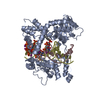
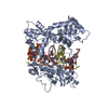
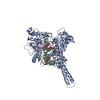
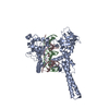
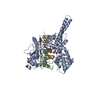
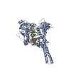
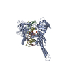
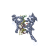
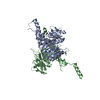
ISOMERASE/DNA / Complex (Isomerase-DNA) / DNA / Topoisomerase I / drug / poison / ISOMERASE-DNA complex
Function / homology
Function and homology information
DNA topoisomerase / DNA topoisomerase type I (single strand cut, ATP-independent) activity / dense fibrillar component / cellular response to luteinizing hormone stimulus / embryonic cleavage / programmed cell death / DNA binding, bending / response to temperature stimulus / supercoiled DNA binding / DNA topological change ...DNA topoisomerase / DNA topoisomerase type I (single strand cut, ATP-independent) activity / dense fibrillar component / cellular response to luteinizing hormone stimulus / embryonic cleavage / programmed cell death / DNA binding, bending / response to temperature stimulus / supercoiled DNA binding / DNA topological change / rRNA transcription / SUMOylation of DNA replication proteins / animal organ regeneration / response to cAMP / male germ cell nucleus / response to gamma radiation / circadian regulation of gene expression / chromosome segregation / molecular condensate scaffold activity / P-body / circadian rhythm / protein-DNA complex / chromatin DNA binding / fibrillar center / single-stranded DNA binding / chromosome / double-stranded DNA binding / perikaryon / DNA replication / RNA polymerase II cis-regulatory region sequence-specific DNA binding / chromatin remodeling / response to xenobiotic stimulus / protein domain specific binding / protein serine/threonine kinase activity / chromatin binding / protein-containing complex binding / nucleolus / DNA binding / RNA binding / nucleoplasm / ATP binding / nucleus Similarity search - Function
Topoisomerase I; Chain A, domain 4 - #10 / : / DNA Topoisomerase I; domain 2 / DNA Topoisomerase I, domain 2 / Yeast DNA topoisomerase - domain 1 / DNA topoisomerase I, DNA binding, eukaryotic-type / DNA topoisomerase I, DNA binding, N-terminal domain 2 / DNA topoisomerase I, DNA binding, N-terminal domain 1 / DNA topoisomerase I, eukaryotic-type / DNA topoisomerase I, catalytic core, alpha/beta subdomain ...Topoisomerase I; Chain A, domain 4 - #10 / : / DNA Topoisomerase I; domain 2 / DNA Topoisomerase I, domain 2 / Yeast DNA topoisomerase - domain 1 / DNA topoisomerase I, DNA binding, eukaryotic-type / DNA topoisomerase I, DNA binding, N-terminal domain 2 / DNA topoisomerase I, DNA binding, N-terminal domain 1 / DNA topoisomerase I, eukaryotic-type / DNA topoisomerase I, catalytic core, alpha/beta subdomain / Topoisomerase I C-terminal domain / DNA topoisomerase I, DNA binding, eukaryotic-type, N-terminal domain superfamily / : / Eukaryotic DNA topoisomerase I, DNA binding fragment / C-terminal topoisomerase domain / DNA Topoisomerase I (eukaryota) / DNA topoisomerase I, active site / Topoisomerase (Topo) IB-type active site signature. / Topoisomerase I; Chain A, domain 3 / Topoisomerase I; Chain A, domain 3 / DNA topoisomerase I / DNA topoisomerase I, catalytic core, eukaryotic-type / DNA topoisomerase I, catalytic core, alpha-helical subdomain, eukaryotic-type / Eukaryotic DNA topoisomerase I, catalytic core / Topoisomerase (Topo) IB-type catalytic domain profile. / DNA breaking-rejoining enzyme, catalytic core / Topoisomerase I; Chain A, domain 4 / Beta Complex / Arc Repressor Mutant, subunit A / Alpha-Beta Complex / Orthogonal Bundle / Mainly Beta / Mainly Alpha / Alpha Beta Similarity search - Domain/homology
In the structure databanks used in Yorodumi, some data are registered as the other names, "COVID-19 virus" and "2019-nCoV". Here are the details of the virus and the list of structure data.
Jan 31, 2019. EMDB accession codes are about to change! (news from PDBe EMDB page)
EMDB accession codes are about to change! (news from PDBe EMDB page)
The allocation of 4 digits for EMDB accession codes will soon come to an end. Whilst these codes will remain in use, new EMDB accession codes will include an additional digit and will expand incrementally as the available range of codes is exhausted. The current 4-digit format prefixed with “EMD-” (i.e. EMD-XXXX) will advance to a 5-digit format (i.e. EMD-XXXXX), and so on. It is currently estimated that the 4-digit codes will be depleted around Spring 2019, at which point the 5-digit format will come into force.
The EM Navigator/Yorodumi systems omit the EMD- prefix.
Related info.:Q: What is EMD? / ID/Accession-code notation in Yorodumi/EM Navigator
Yorodumi is a browser for structure data from EMDB, PDB, SASBDB, etc.
This page is also the successor to EM Navigator detail page, and also detail information page/front-end page for Omokage search.
The word "yorodu" (or yorozu) is an old Japanese word meaning "ten thousand". "mi" (miru) is to see.
Related info.:EMDB / PDB / SASBDB / Comparison of 3 databanks / Yorodumi Search / Aug 31, 2016. New EM Navigator & Yorodumi / Yorodumi Papers / Jmol/JSmol / Function and homology information / Changes in new EM Navigator and Yorodumi
 Movie
Movie Controller

 Yorodumi
 Open data
 Basic information
 Components
 Keywords
 Function and homology information
 Homo sapiens (human)
 X-RAY DIFFRACTION /
  Authors
 Citation
 Structure visualization
 Downloads & links
 Other downloads
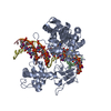


 PDBj
PDBj


 Assembly
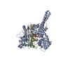

 Spodoptera frugiperda (fall armyworm) / References: UniProt: P11387, EC: 5.99.1.2


 Mass: 439.461 Da / Num. of mol.: 1 / Source method: obtained synthetically / Formula: C23H25N3O6
 Mass: 421.446 Da / Num. of mol.: 1 / Source method: obtained synthetically / Formula: C23H23N3O5
 Mass: 200.590 Da / Num. of mol.: 1 / Source method: obtained synthetically / Formula: Hg
 Mass: 194.226 Da / Num. of mol.: 1 / Source method: obtained synthetically / Formula: C8H18O5 / Comment: precipitant*YM
 Sample preparation
 / Beamline: X25
 Processing