 Movie
Movie Controller

[English] 日本語
 Yorodumi
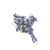
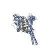
- PDB-1t8i: Human DNA Topoisomerase I (70 Kda) In Complex With The Poison Cam... -
+ Open data

- Basic information
| Entry | Database: PDB / ID: 1t8i | ||||||
|---|---|---|---|---|---|---|---|
| Title | Human DNA Topoisomerase I (70 Kda) In Complex With The Poison Camptothecin and Covalent Complex With A 22 Base Pair DNA Duplex | ||||||
 Components Components |
| ||||||
 Keywords Keywords | ISOMERASE/DNA / COMPLEX (ISOMERASE-DNA) / DNA / TOPOISOMERASE I / DRUG / POISON / ISOMERASE-DNA complex | ||||||
| Function / homology |  Function and homology information Function and homology informationDNA topoisomerase / DNA topoisomerase type I (single strand cut, ATP-independent) activity / dense fibrillar component / cellular response to luteinizing hormone stimulus / programmed cell death / DNA binding, bending / supercoiled DNA binding / response to temperature stimulus / DNA topological change / animal organ regeneration ...DNA topoisomerase / DNA topoisomerase type I (single strand cut, ATP-independent) activity / dense fibrillar component / cellular response to luteinizing hormone stimulus / programmed cell death / DNA binding, bending / supercoiled DNA binding / response to temperature stimulus / DNA topological change / animal organ regeneration / rRNA transcription / SUMOylation of DNA replication proteins / response to cAMP / response to gamma radiation / circadian regulation of gene expression / chromosome segregation / molecular condensate scaffold activity / circadian rhythm / P-body / protein-DNA complex / chromatin DNA binding / fibrillar center / single-stranded DNA binding / chromosome / double-stranded DNA binding / perikaryon / DNA replication / response to xenobiotic stimulus / RNA polymerase II cis-regulatory region sequence-specific DNA binding / chromatin remodeling / protein domain specific binding / protein serine/threonine kinase activity / chromatin binding / nucleolus / protein-containing complex binding / DNA binding / RNA binding / nucleoplasm / ATP binding / nucleus Similarity search - Function | ||||||
| Biological species |  Homo sapiens (human) Homo sapiens (human) | ||||||
| Method |  X-RAY DIFFRACTION / SYNCHROTRON / MOLECULAR REPLACEMENT / Resolution: 3 Å X-RAY DIFFRACTION / SYNCHROTRON / MOLECULAR REPLACEMENT / Resolution: 3 Å | ||||||
 Authors Authors | Staker, B.L. / Feese, M.D. / Cushman, M. / Pommier, Y. / Zembower, D. / Stewart, L. / Burgin, A.B. | ||||||
 Citation Citation | Journal: J.Med.Chem. / Year: 2005 Title: Structures of three classes of anticancer agents bound to the human topoisomerase I-DNA covalent complex Authors: Staker, B.L. / Feese, M.D. / Cushman, M. / Pommier, Y. / Zembower, D. / Stewart, L. / Burgin, A.B. | ||||||
| History |
| ||||||
| Remark 600 | HETEROGEN Atom O3* of thymine 10, chain B is missing due to the covalent bond formed between ...HETEROGEN Atom O3* of thymine 10, chain B is missing due to the covalent bond formed between thymine 10 and phosphotyrosine (PTR) 723 |
- Structure visualization
| Structure viewer | Molecule: MolmilJmol/JSmol |
|---|
- Downloads & links
-Download
| PDBx/mmCIF format | 1t8i.cif.gz | 157.1 KB | Display | PDBx/mmCIF format |
|---|---|---|---|---|
| PDB format | pdb1t8i.ent.gz | 117.8 KB | Display | PDB format |
| PDBx/mmJSON format | 1t8i.json.gz | Tree view | PDBx/mmJSON format | |
| Others |  Other downloads Other downloads |
-Validation report
| Arichive directory | https://data.pdbj.org/pub/pdb/validation_reports/t8/1t8iftp://data.pdbj.org/pub/pdb/validation_reports/t8/1t8i | HTTPS FTP |
|---|
-Related structure data
| Related structure data | 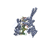 1sc7C  1seuC 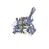 1k4tS S: Starting model for refinement C: citing same article ( |
|---|---|
| Similar structure data |


-Links
 PDBj
PDBj


- Assembly
| Deposited unit | 
| ||||||||
|---|---|---|---|---|---|---|---|---|---|
| 1 |
| ||||||||
| Unit cell |
|
-Components
| #1: DNA chain | Mass: 3061.057 Da / Num. of mol.: 1 / Source method: obtained synthetically |
|---|---|
| #2: DNA chain | Mass: 3716.518 Da / Num. of mol.: 1 / Source method: obtained synthetically |
| #3: DNA chain | Mass: 6690.362 Da / Num. of mol.: 1 / Source method: obtained synthetically |
| #4: Protein | Mass: 70248.023 Da / Num. of mol.: 1 Source method: isolated from a genetically manipulated source Source: (gene. exp.) Homo sapiens (human) / Gene: TOP1 / Production host:   Spodoptera frugiperda (fall armyworm) / References: UniProt: P11387, EC: 5.99.1.2 Spodoptera frugiperda (fall armyworm) / References: UniProt: P11387, EC: 5.99.1.2 |
| #5: Chemical | ChemComp-EHD / 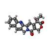  Mass: 348.352 Da / Num. of mol.: 1 / Source method: obtained synthetically / Formula: C20H16N2O4 / Comment: inhibitor*YM Mass: 348.352 Da / Num. of mol.: 1 / Source method: obtained synthetically / Formula: C20H16N2O4 / Comment: inhibitor*YM |
| Has protein modification | Y |
-Experimental details
-Experiment
| Experiment | Method: X-RAY DIFFRACTION / Number of used crystals: 1 |
|---|
- Sample preparation
| Crystal | Density Matthews: 2.9 Å3/Da / Density % sol: 57.59 % | ||||||||||||||||||||||||
|---|---|---|---|---|---|---|---|---|---|---|---|---|---|---|---|---|---|---|---|---|---|---|---|---|---|
| Crystal grow | Temperature: 289 K / Method: vapor diffusion, sitting drop / pH: 6.4 Details: PEG 8000, ammonium sulfate, MES, pH 6.4, VAPOR DIFFUSION, SITTING DROP, temperature 289K | ||||||||||||||||||||||||
| Components of the solutions |
|
-Data collection
| Diffraction | Mean temperature: 100 K |
|---|---|
| Diffraction source | Source: SYNCHROTRON / Site: APS  / Beamline: 32-ID / Beamline: 32-ID |
| Radiation | Protocol: SINGLE WAVELENGTH / Monochromatic (M) / Laue (L): M / Scattering type: x-ray |
| Radiation wavelength | Relative weight: 1 |
| Reflection | Resolution: 3→20 Å / Num. all: 16492 / Num. obs: 16492 / % possible obs: 91 % / Observed criterion σ(F): 0 / Observed criterion σ(I): 0 / Rsym value: 0.08 / Net I/σ(I): 13.7 |
| Reflection shell | Resolution: 3→3.16 Å / Mean I/σ(I) obs: 1.7 / Num. unique all: 1427 / Rsym value: 0.427 / % possible all: 47 |
- Processing
| Software |
| ||||||||||||||||||||||||||||||||||||||||||||||||||||||||||||||||||||||||||||||||||||||||||||||||||||||||||||||||||||||||||||||||||||||||||||||||||||||||||||||||
|---|---|---|---|---|---|---|---|---|---|---|---|---|---|---|---|---|---|---|---|---|---|---|---|---|---|---|---|---|---|---|---|---|---|---|---|---|---|---|---|---|---|---|---|---|---|---|---|---|---|---|---|---|---|---|---|---|---|---|---|---|---|---|---|---|---|---|---|---|---|---|---|---|---|---|---|---|---|---|---|---|---|---|---|---|---|---|---|---|---|---|---|---|---|---|---|---|---|---|---|---|---|---|---|---|---|---|---|---|---|---|---|---|---|---|---|---|---|---|---|---|---|---|---|---|---|---|---|---|---|---|---|---|---|---|---|---|---|---|---|---|---|---|---|---|---|---|---|---|---|---|---|---|---|---|---|---|---|---|---|---|---|
| Refinement | Method to determine structure: MOLECULAR REPLACEMENT Starting model: 1K4T Resolution: 3→20 Å / Cor.coef. Fo:Fc: 0.913 / Cor.coef. Fo:Fc free: 0.862 / SU B: 24.369 / SU ML: 0.456 / Cross valid method: THROUGHOUT / ESU R Free: 0.536 / Stereochemistry target values: MAXIMUM LIKELIHOOD / Details: HYDROGENS HAVE BEEN ADDED IN THE RIDING POSITIONS
| ||||||||||||||||||||||||||||||||||||||||||||||||||||||||||||||||||||||||||||||||||||||||||||||||||||||||||||||||||||||||||||||||||||||||||||||||||||||||||||||||
| Solvent computation | Ion probe radii: 0.8 Å / Shrinkage radii: 0.8 Å / VDW probe radii: 1.4 Å / Solvent model: BABINET MODEL WITH MASK | ||||||||||||||||||||||||||||||||||||||||||||||||||||||||||||||||||||||||||||||||||||||||||||||||||||||||||||||||||||||||||||||||||||||||||||||||||||||||||||||||
| Displacement parameters | Biso mean: 72.185 Å2
| ||||||||||||||||||||||||||||||||||||||||||||||||||||||||||||||||||||||||||||||||||||||||||||||||||||||||||||||||||||||||||||||||||||||||||||||||||||||||||||||||
| Refinement step | Cycle: LAST / Resolution: 3→20 Å
| ||||||||||||||||||||||||||||||||||||||||||||||||||||||||||||||||||||||||||||||||||||||||||||||||||||||||||||||||||||||||||||||||||||||||||||||||||||||||||||||||
| Refine LS restraints |
| ||||||||||||||||||||||||||||||||||||||||||||||||||||||||||||||||||||||||||||||||||||||||||||||||||||||||||||||||||||||||||||||||||||||||||||||||||||||||||||||||
| LS refinement shell | Resolution: 3→3.158 Å / Total num. of bins used: 10 /
|
