[English] 日本語
Yorodumi
- PDB-1j7p: Solution structure of Calcium calmodulin C-terminal domain -

+
Open data


ID or keywords:

Loading...

-
Basic information

Entry
Database: PDB / ID: 1j7p
TitleSolution structure of Calcium calmodulin C-terminal domain
ComponentsCalmodulin
KeywordsMETAL BINDING PROTEIN / EF hands / helix bundle / calcium / dipolar coupling
Function / homology
Function and homology information


: / establishment of protein localization to mitochondrial membrane / type 3 metabotropic glutamate receptor binding / CaM pathway / Cam-PDE 1 activation / Sodium/Calcium exchangers / Calmodulin induced events / regulation of synaptic vesicle endocytosis / Reduction of cytosolic Ca++ levels / CREB1 phosphorylation through the activation of CaMKII/CaMKK/CaMKIV cascasde ...: / establishment of protein localization to mitochondrial membrane / type 3 metabotropic glutamate receptor binding / CaM pathway / Cam-PDE 1 activation / Sodium/Calcium exchangers / Calmodulin induced events / regulation of synaptic vesicle endocytosis / Reduction of cytosolic Ca++ levels / CREB1 phosphorylation through the activation of CaMKII/CaMKK/CaMKIV cascasde / Activation of Ca-permeable Kainate Receptor / Loss of phosphorylation of MECP2 at T308 / CREB1 phosphorylation through the activation of Adenylate Cyclase / PKA activation / negative regulation of high voltage-gated calcium channel activity / CaMK IV-mediated phosphorylation of CREB / Glycogen breakdown (glycogenolysis) / positive regulation of cyclic-nucleotide phosphodiesterase activity / regulation of synaptic vesicle exocytosis / organelle localization by membrane tethering / negative regulation of calcium ion export across plasma membrane / CLEC7A (Dectin-1) induces NFAT activation / mitochondrion-endoplasmic reticulum membrane tethering / autophagosome membrane docking / regulation of cardiac muscle cell action potential / Activation of RAC1 downstream of NMDARs / response to corticosterone / positive regulation of DNA binding / positive regulation of ryanodine-sensitive calcium-release channel activity / nitric-oxide synthase binding / regulation of cell communication by electrical coupling involved in cardiac conduction / Negative regulation of NMDA receptor-mediated neuronal transmission / negative regulation of peptidyl-threonine phosphorylation / Synthesis of IP3 and IP4 in the cytosol / Unblocking of NMDA receptors, glutamate binding and activation / Phase 0 - rapid depolarisation / negative regulation of ryanodine-sensitive calcium-release channel activity / protein phosphatase activator activity / RHO GTPases activate PAKs / : / Ion transport by P-type ATPases / Long-term potentiation / Uptake and function of anthrax toxins / Regulation of MECP2 expression and activity / Calcineurin activates NFAT / adenylate cyclase binding / catalytic complex / DARPP-32 events / detection of calcium ion / regulation of cardiac muscle contraction / regulation of ryanodine-sensitive calcium-release channel activity / Smooth Muscle Contraction / cellular response to interferon-beta / RHO GTPases activate IQGAPs / regulation of cardiac muscle contraction by regulation of the release of sequestered calcium ion / calcium channel inhibitor activity / Protein methylation / phosphatidylinositol 3-kinase binding / eNOS activation / enzyme regulator activity / activation of adenylate cyclase activity / regulation of release of sequestered calcium ion into cytosol by sarcoplasmic reticulum / Activation of AMPK downstream of NMDARs / Tetrahydrobiopterin (BH4) synthesis, recycling, salvage and regulation / : / Ion homeostasis / titin binding / regulation of calcium-mediated signaling / positive regulation of protein autophosphorylation / voltage-gated potassium channel complex / sperm midpiece / calcium channel complex / response to amphetamine / substantia nigra development / adenylate cyclase activator activity / Ras activation upon Ca2+ influx through NMDA receptor / nitric-oxide synthase regulator activity / regulation of heart rate / sarcomere / FCERI mediated Ca+2 mobilization / protein serine/threonine kinase activator activity / FCGR3A-mediated IL10 synthesis / VEGFR2 mediated vascular permeability / Antigen activates B Cell Receptor (BCR) leading to generation of second messengers / VEGFR2 mediated cell proliferation / regulation of cytokinesis / positive regulation of peptidyl-threonine phosphorylation / positive regulation of nitric-oxide synthase activity / Translocation of SLC2A4 (GLUT4) to the plasma membrane / spindle microtubule / RAF activation / positive regulation of receptor signaling pathway via JAK-STAT / response to calcium ion / Transcriptional activation of mitochondrial biogenesis / positive regulation of protein serine/threonine kinase activity / mitochondrial membrane / Stimuli-sensing channels / cellular response to type II interferon / G2/M transition of mitotic cell cycle / spindle pole
Similarity search - Function
: / EF-hand / Recoverin; domain 1 / EF-hand domain pair / EF-hand, calcium binding motif / EF-Hand 1, calcium-binding site / EF-hand calcium-binding domain. / EF-hand calcium-binding domain profile. / EF-hand domain / EF-hand domain pair ...: / EF-hand / Recoverin; domain 1 / EF-hand domain pair / EF-hand, calcium binding motif / EF-Hand 1, calcium-binding site / EF-hand calcium-binding domain. / EF-hand calcium-binding domain profile. / EF-hand domain / EF-hand domain pair / Orthogonal Bundle / Mainly Alpha
Similarity search - Domain/homology
Calmodulin-1 / Calmodulin-3
Similarity search - Component
Biological speciesHomo sapiens (human)
MethodSOLUTION NMR / simulated annealing
AuthorsChou, J.J. / Klee, C.B. / Bax, A.
CitationJournal: Nat.Struct.Biol. / Year: 2001
Title: Solution structure of Ca(2+)-calmodulin reveals flexible hand-like properties of its domains.
Authors: Chou, J.J. / Li, S. / Klee, C.B. / Bax, A.
History
DepositionMay 17, 2001Deposition site: RCSB / Processing site: RCSB
Revision 1.0Nov 7, 2001Provider: repository / Type: Initial release
Revision 1.1Apr 27, 2008Group: Version format compliance
Revision 1.2Jul 13, 2011Group: Version format compliance
Revision 1.3Feb 23, 2022Group: Data collection / Database references / Derived calculations
Category: database_2 / pdbx_nmr_spectrometer ...database_2 / pdbx_nmr_spectrometer / pdbx_struct_assembly / pdbx_struct_conn_angle / pdbx_struct_oper_list / struct_conn / struct_site
Item: _database_2.pdbx_DOI / _database_2.pdbx_database_accession ..._database_2.pdbx_DOI / _database_2.pdbx_database_accession / _pdbx_nmr_spectrometer.model / _pdbx_struct_conn_angle.ptnr1_auth_comp_id / _pdbx_struct_conn_angle.ptnr1_auth_seq_id / _pdbx_struct_conn_angle.ptnr1_label_atom_id / _pdbx_struct_conn_angle.ptnr1_label_comp_id / _pdbx_struct_conn_angle.ptnr1_label_seq_id / _pdbx_struct_conn_angle.ptnr3_auth_comp_id / _pdbx_struct_conn_angle.ptnr3_auth_seq_id / _pdbx_struct_conn_angle.ptnr3_label_atom_id / _pdbx_struct_conn_angle.ptnr3_label_comp_id / _pdbx_struct_conn_angle.ptnr3_label_seq_id / _pdbx_struct_conn_angle.value / _struct_conn.pdbx_dist_value / _struct_conn.ptnr1_auth_comp_id / _struct_conn.ptnr1_auth_seq_id / _struct_conn.ptnr1_label_asym_id / _struct_conn.ptnr1_label_atom_id / _struct_conn.ptnr1_label_comp_id / _struct_conn.ptnr1_label_seq_id / _struct_conn.ptnr2_auth_comp_id / _struct_conn.ptnr2_auth_seq_id / _struct_conn.ptnr2_label_asym_id / _struct_conn.ptnr2_label_atom_id / _struct_conn.ptnr2_label_comp_id / _struct_conn.ptnr2_label_seq_id / _struct_site.pdbx_auth_asym_id / _struct_site.pdbx_auth_comp_id / _struct_site.pdbx_auth_seq_id
Revision 1.4May 22, 2024Group: Data collection / Category: chem_comp_atom / chem_comp_bond

-
Structure visualization

Structure viewerMolecule:
MolmilJmol/JSmol

Downloads & links

-
Assembly

Deposited unit
A: Calmodulin
hetero molecules


Theoretical massNumber of molelcules
Total (without water)7,8183
Polymers7,7371
Non-polymers802
Water00
1


  • Idetical with deposited unit
  • defined by author
TypeNameSymmetry operationNumber
identity operation1_555x,y,z1
NMR ensembles
DataCriteria
Number of conformers (submitted / calculated)3 / 3structure with the lowest dipolar energy
RepresentativeModel #2lowest dipolar energy

-
Components

#1: Protein Calmodulin


Mass: 7737.468 Da / Num. of mol.: 1 / Fragment: C-terminal domain
Source method: isolated from a genetically manipulated source
Source: (gene. exp.) Homo sapiens (human) / Production host: Escherichia coli (E. coli) / Strain (production host): AR58 / References: UniProt: P62158, UniProt: P0DP23*PLUS
#2: Chemical ChemComp-CA / CALCIUM ION


Mass: 40.078 Da / Num. of mol.: 2 / Source method: obtained synthetically / Formula: Ca

-
Experimental details

-
Experiment

ExperimentMethod: SOLUTION NMR
NMR experiment
Conditions-IDExperiment-IDSolution-IDType
1113D HNCO (NH coupled)
1213D HNCO (C'CA coupled)
131CBCA(CO)NH (Quantitative J)
141TROSY HNCO (Quantitative J)
151HN(CO)CA (C'HA coupled)
1613D 13C-separated NOESY
NMR detailsText: A total of five sets of dipolar couplings are measured, including the one-bond NH, CAHA, C'CA, and NC' couplings, and the two-bond C'HA couplings. Additionally, the CBHB dipolar couplings were ...Text: A total of five sets of dipolar couplings are measured, including the one-bond NH, CAHA, C'CA, and NC' couplings, and the two-bond C'HA couplings. Additionally, the CBHB dipolar couplings were measure to assign chi-1 rotamers for locked sidechains.

-
Sample preparation

Details
Solution-IDContentsSolvent system
11mM calmodulin U-15N,13C; 100mM KCl, 16mM CaCl2, pH 7.0;95% H2O/5% D2O
21mM calmodulin U-15N,13C; 10mM KCl, 16mM CaCl2, pH 7.0; 15 mg/ml Pf1;95% H2O/5% D2O
30.5mM calmodulin U-15N,13C; 100mM KCl, 6mM CaCl2, pH 7.0; 18 mg/ml Pf1;95% H2O/5% D2O
Sample conditions
Conditions-IDIonic strengthpHPressure (kPa)Temperature (K)
1100mM KCl 7ambient 305 K
210mM KCl 7ambient 305 K
3100mM KCl 7ambient 305 K
Crystal grow
*PLUS
Method: other / Details: NMR

-
NMR measurement

RadiationProtocol: SINGLE WAVELENGTH / Monochromatic (M) / Laue (L): M
Radiation wavelengthRelative weight: 1
NMR spectrometer
TypeManufacturerModelField strength (MHz)Spectrometer-ID
Bruker AVANCEBrukerAVANCE5001
Bruker AVANCEBrukerAVANCE6002
Bruker AVANCEBrukerAVANCE7503

-
Processing

NMR software
NameVersionDeveloperClassification
X-PLOR3.851Brungerrefinement
NMRPipe2Delaglioprocessing
Refinement
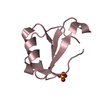
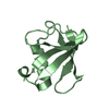
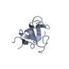
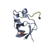
Method: simulated annealing / Software ordinal: 1
Details: This structure is determined mainly by residual dipolar couplings measured in A liquid crystalline Pf1 medium. The structure calculation scheme, described in the paper, is based on the idea ...Details: This structure is determined mainly by residual dipolar couplings measured in A liquid crystalline Pf1 medium. The structure calculation scheme, described in the paper, is based on the idea of refining existing structural models against dipolar couplings to derive the correct structure. Here a total of 305 backbone dipolar couplings are used to refine the backbone structure. Additionally, 35 sidechain dipolar couplings and 81 3-bond J couplings are used to determine the sidechain chi1 and chi2 rotamers as well as the presence of rotameric averaging. A total of three structures (model 1-3) were calculated starting from the 1. A crystal structure of Ca-calmodulin (PDB entry 1EXR), the NMR structure of apo-calmodulin (1F70), and the crystal structure of Ca-ligated parvalbumin (1CDP). The convergence of refinement is indicated by the small average RMSD between the three calculated structures and the average coordinates (0.26 A for backbone and 0.90 for all heavy atoms). During the three-stage simulated annealing described in the paper, restraints are included for most previously established hydrogen bonds, but have only minute effects (< 0.3 A) on the final structure.
NMR representativeSelection criteria: lowest dipolar energy
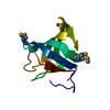
NMR ensembleConformer selection criteria: structure with the lowest dipolar energy
Conformers calculated total number: 3 / Conformers submitted total number: 3

+
About Yorodumi

-
News

-
Feb 9, 2022. New format data for meta-information of EMDB entries

New format data for meta-information of EMDB entries

  • Version 3 of the EMDB header file is now the official format.
  • The previous official version 1.9 will be removed from the archive.

Related info.:EMDB header

External links:wwPDB to switch to version 3 of the EMDB data model

-
Aug 12, 2020. Covid-19 info

Covid-19 info

URL: https://pdbjlvh1.pdbj.org/emnavi/covid19.php

New page: Covid-19 featured information page in EM Navigator.

Related info.:Covid-19 info / Mar 5, 2020. Novel coronavirus structure data

+
Mar 5, 2020. Novel coronavirus structure data

Novel coronavirus structure data

Related info.:Yorodumi Speices / Aug 12, 2020. Covid-19 info

External links:COVID-19 featured content - PDBj / Molecule of the Month (242):Coronavirus Proteases

+
Jan 31, 2019. EMDB accession codes are about to change! (news from PDBe EMDB page)

EMDB accession codes are about to change! (news from PDBe EMDB page)

  • The allocation of 4 digits for EMDB accession codes will soon come to an end. Whilst these codes will remain in use, new EMDB accession codes will include an additional digit and will expand incrementally as the available range of codes is exhausted. The current 4-digit format prefixed with “EMD-” (i.e. EMD-XXXX) will advance to a 5-digit format (i.e. EMD-XXXXX), and so on. It is currently estimated that the 4-digit codes will be depleted around Spring 2019, at which point the 5-digit format will come into force.
  • The EM Navigator/Yorodumi systems omit the EMD- prefix.

Related info.:Q: What is EMD? / ID/Accession-code notation in Yorodumi/EM Navigator

External links:EMDB Accession Codes are Changing Soon! / Contact to PDBj

+
Jul 12, 2017. Major update of PDB

Major update of PDB

  • wwPDB released updated PDB data conforming to the new PDBx/mmCIF dictionary.
  • This is a major update changing the version number from 4 to 5, and with Remediation, in which all the entries are updated.
  • In this update, many items about electron microscopy experimental information are reorganized (e.g. em_software).
  • Now, EM Navigator and Yorodumi are based on the updated data.

External links:wwPDB Remediation / Enriched Model Files Conforming to OneDep Data Standards Now Available in the PDB FTP Archive

-
Yorodumi

Thousand views of thousand structures

  • Yorodumi is a browser for structure data from EMDB, PDB, SASBDB, etc.
  • This page is also the successor to EM Navigator detail page, and also detail information page/front-end page for Omokage search.
  • The word "yorodu" (or yorozu) is an old Japanese word meaning "ten thousand". "mi" (miru) is to see.

Related info.:EMDB / PDB / SASBDB / Comparison of 3 databanks / Yorodumi Search / Aug 31, 2016. New EM Navigator & Yorodumi / Yorodumi Papers / Jmol/JSmol / Function and homology information / Changes in new EM Navigator and Yorodumi

Read more