Mass: 197.382 Da / Num. of mol.: 1 / Source method: obtained synthetically / Formula: C2HBrClF3
-
Experimental details
-
Experiment
Experiment
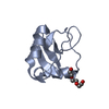
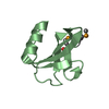
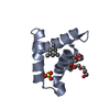
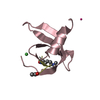
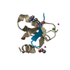
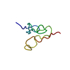
Method: SOLUTION NMR Details: The mechanism(s) of volatile anesthetic effects are poorly understood. We examined whether VA binding to druggable sites in calmodulin would effect [Ca2+]4-CaM dependent activity of enzymes. ...Details: The mechanism(s) of volatile anesthetic effects are poorly understood. We examined whether VA binding to druggable sites in calmodulin would effect [Ca2+]4-CaM dependent activity of enzymes. We used high resolution NMR spectroscopy to determine the structure of the halothane [Ca2+]4-CaM complex, determining that the halothane molecules bind in the druggable sites. We used fluorescence assays to determine that VA mediate [Ca2+]4-CaM activation of smMLCK, but not the kd of [Ca2+]4-CaM binding to skMLCK. These results suggest that VA do not mediate [Ca2+]4-CaM dependent MLCK activity via direct interactions with druggable sites on [Ca2+]4-CaM.
NMR experiment
Conditions-ID
Experiment-ID
Solution-ID
Type
1
1
1
3D HNCO
1
2
1
3D HNCA
1
3
1
3D HN(CA)CB
1
4
1
3DHBHA(CO)NH
1
5
1
3DH(CCO)NH
1
6
1
3D (H)CCH-TOCSY
1
7
1
3D (H)CCH-COSY
1
8
1
3D 1H-15N NOESY
1
9
1
3D 1H-13C NOESY
1
10
2
3D HNHA
1
11
2
2D 1H-15N NOE
-
Sample preparation
Details
Solution-ID
Contents
Solvent system
1
2 mM [U-99% 13C; U-99% 15N] CALCIUM ION, 20 mM CALCIUM ION, 20 mM N-{[2-({[1-(4-CARBOXYBUTANOYL)AMINO]-2-PHENYLETHYL}-HYDROXYPHOSPHINYL)OXY]ACETYL}-2-PHENYLETHYLAMINE, 95% H2O/5% D2O
95% H2O/5% D2O
2
2 mM [U-99% 15N] CALCIUM ION, 20 mM CALCIUM ION, 20 mM N-{[2-({[1-(4-CARBOXYBUTANOYL)AMINO]-2-PHENYLETHYL}-HYDROXYPHOSPHINYL)OXY]ACETYL}-2-PHENYLETHYLAMINE, 95% H2O/5% D2O
Method: simulated annealing, CHARMm22 energy minimization / Software ordinal: 1 Details: XPLOR, 100 steps steepest descend - final refinement
NMR constraints
NOE constraints total: 808 / NOE intraresidue total count: 337 / NOE long range total count: 137 / NOE medium range total count: 137 / NOE sequential total count: 188 / Hydrogen bond constraints total count: 46 / Protein chi angle constraints total count: 65 / Protein other angle constraints total count: 65 / Protein phi angle constraints total count: 65 / Protein psi angle constraints total count: 65
NMR representative
Selection criteria: closest to the average
NMR ensemble
Conformer selection criteria: structures with the lowest energy Conformers calculated total number: 50 / Conformers submitted total number: 5
+
About Yorodumi
-
News
-
Feb 9, 2022. New format data for meta-information of EMDB entries
New format data for meta-information of EMDB entries
Version 3 of the EMDB header file is now the official format.
The previous official version 1.9 will be removed from the archive.
In the structure databanks used in Yorodumi, some data are registered as the other names, "COVID-19 virus" and "2019-nCoV". Here are the details of the virus and the list of structure data.
Jan 31, 2019. EMDB accession codes are about to change! (news from PDBe EMDB page)
EMDB accession codes are about to change! (news from PDBe EMDB page)
The allocation of 4 digits for EMDB accession codes will soon come to an end. Whilst these codes will remain in use, new EMDB accession codes will include an additional digit and will expand incrementally as the available range of codes is exhausted. The current 4-digit format prefixed with “EMD-” (i.e. EMD-XXXX) will advance to a 5-digit format (i.e. EMD-XXXXX), and so on. It is currently estimated that the 4-digit codes will be depleted around Spring 2019, at which point the 5-digit format will come into force.
The EM Navigator/Yorodumi systems omit the EMD- prefix.
Related info.:Q: What is EMD? / ID/Accession-code notation in Yorodumi/EM Navigator
Yorodumi is a browser for structure data from EMDB, PDB, SASBDB, etc.
This page is also the successor to EM Navigator detail page, and also detail information page/front-end page for Omokage search.
The word "yorodu" (or yorozu) is an old Japanese word meaning "ten thousand". "mi" (miru) is to see.
Related info.:EMDB / PDB / SASBDB / Comparison of 3 databanks / Yorodumi Search / Aug 31, 2016. New EM Navigator & Yorodumi / Yorodumi Papers / Jmol/JSmol / Function and homology information / Changes in new EM Navigator and Yorodumi
 Movie
Movie Controller

 Yorodumi
 Open data
 Basic information
 Components
 Keywords
 Function and homology information
 Homo sapiens (human)
 Authors
 Citation
 Structure visualization
 Downloads & links
 Other downloads
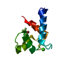


 PDBj
PDBj


 Assembly


 Mass: 40.078 Da / Num. of mol.: 2 / Source method: obtained synthetically / Formula: Ca
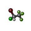
 Mass: 197.382 Da / Num. of mol.: 1 / Source method: obtained synthetically / Formula: C2HBrClF3
 HNCA
HNCA Sample preparation
 Processing