 Movie
Movie Controller

[English] 日本語
 Yorodumi
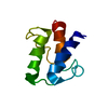
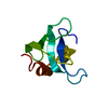
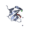
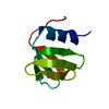
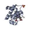
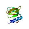
- PDB-2zdj: Crystal Structure of TTMA177, a Hypothetical Protein from Thermus... -
+ Open data

- Basic information
| Entry | Database: PDB / ID: 2zdj | ||||||
|---|---|---|---|---|---|---|---|
| Title | Crystal Structure of TTMA177, a Hypothetical Protein from Thermus thermophilus phage TMA | ||||||
 Components Components | hypothetical protein TTMA177 | ||||||
 Keywords Keywords | STRUCTURAL GENOMICS / UNKNOWN FUNCTION / Alpha and beta proteins (a+b) / Cystatin-like / NPPSFA / National Project on Protein Structural and Functional Analyses / RIKEN Structural Genomics/Proteomics Initiative / RSGI | ||||||
| Function / homology | Nuclear Transport Factor 2; Chain: A, - #450 / : / : / Domain of unknown function (DUF6839) / Nuclear Transport Factor 2; Chain: A, / Roll / Alpha Beta / Hypothetical protein TTMA177 Function and homology information Function and homology information | ||||||
| Biological species | Thermus thermophilus phage TMA (others) | ||||||
| Method |  X-RAY DIFFRACTION / SYNCHROTRON / SAD / Resolution: 2.2 Å X-RAY DIFFRACTION / SYNCHROTRON / SAD / Resolution: 2.2 Å | ||||||
 Authors Authors | Agari, Y. / Tamakoshi, M. / Yamagishi, A. / Shinkai, A. / Ebihara, A. / Yokoyama, S. / Kuramitsu, S. / Oshima, T. / RIKEN Structural Genomics/Proteomics Initiative (RSGI) | ||||||
 Citation Citation | Journal: To be Published Title: Crystal Structure of TTMA177, a Hypothetical Protein from Thermus thermophilus phage TMA Authors: Agari, Y. / Tamakoshi, M. / Yamagishi, A. / Shinkai, A. / Ebihara, A. / Yokoyama, S. / Kuramitsu, S. / Oshima, T. | ||||||
| History |
|
- Structure visualization
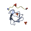
| Structure viewer | Molecule: MolmilJmol/JSmol |
|---|
- Downloads & links
-Download
| PDBx/mmCIF format | 2zdj.cif.gz | 69.3 KB | Display | PDBx/mmCIF format |
|---|---|---|---|---|
| PDB format | pdb2zdj.ent.gz | 52.6 KB | Display | PDB format |
| PDBx/mmJSON format | 2zdj.json.gz | Tree view | PDBx/mmJSON format | |
| Others |  Other downloads Other downloads |
-Validation report
| Arichive directory | https://data.pdbj.org/pub/pdb/validation_reports/zd/2zdjftp://data.pdbj.org/pub/pdb/validation_reports/zd/2zdj | HTTPS FTP |
|---|
-Related structure data
| Similar structure data | |
|---|---|
| Other databases |


-Links
 PDBj
PDBj- Assembly
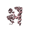
| Deposited unit | 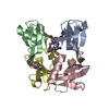
| ||||||||
|---|---|---|---|---|---|---|---|---|---|
| 1 | 
| ||||||||
| 2 | 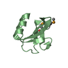
| ||||||||
| 3 | 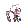
| ||||||||
| 4 | 
| ||||||||
| Unit cell |
|
-Components
| #1: Protein | Mass: 8182.890 Da / Num. of mol.: 4 / Fragment: Residues 1-69 Source method: isolated from a genetically manipulated source Source: (gene. exp.) Thermus thermophilus phage TMA (others) Plasmid: pET-21a(+) / Production host:  #2: Water | ChemComp-HOH / |  Mass: 18.015 Da / Num. of mol.: 109 / Source method: isolated from a natural source / Formula: H2O Mass: 18.015 Da / Num. of mol.: 109 / Source method: isolated from a natural source / Formula: H2OHas protein modification | Y | Sequence details | THERE IS NO UNP REFERENCE SEQUENCE DATABASE FOR THIS PROTEIN AT THE TIME OF PROCESSING | |
|---|
-Experimental details
-Experiment
| Experiment | Method: X-RAY DIFFRACTION / Number of used crystals: 1 |
|---|
- Sample preparation
| Crystal | Density Matthews: 2.22 Å3/Da / Density % sol: 44.6 % |
|---|---|
| Crystal grow | Temperature: 293 K / Method: vapor diffusion, hanging drop / pH: 6 Details: 15% (w/v) PEG 8000, 75mM MES, 150mM Calcium Acetate, pH 6.0, VAPOR DIFFUSION, HANGING DROP, temperature 293K |
-Data collection
| Diffraction | Mean temperature: 100 K |
|---|---|
| Diffraction source | Source: SYNCHROTRON / Site: SPring-8  / Beamline: BL26B2 / Wavelength: 0.97893 Å / Beamline: BL26B2 / Wavelength: 0.97893 Å |
| Detector | Type: MARMOSAIC 225 mm CCD / Detector: CCD / Date: Sep 22, 2007 Details: A fixed exit Si double crystal monochromator followed by a two dimensional focusing mirror which is coated in rhodium. |
| Radiation | Monochromator: Fixed exit Si double crystal monochromator / Protocol: SINGLE WAVELENGTH / Monochromatic (M) / Laue (L): M / Scattering type: x-ray |
| Radiation wavelength | Wavelength: 0.97893 Å / Relative weight: 1 |
| Reflection | Resolution: 2.2→50 Å / Num. all: 15444 / Num. obs: 15444 / % possible obs: 100 % / Redundancy: 10.1 % / Biso Wilson estimate: 24.6 Å2 / Rmerge(I) obs: 0.08 / Net I/σ(I): 31.44 |
| Reflection shell | Resolution: 2.2→2.28 Å / Redundancy: 10.4 % / Rmerge(I) obs: 0.292 / Mean I/σ(I) obs: 8.125 / Num. unique all: 1508 / % possible all: 100 |
- Processing
| Software |
| ||||||||||||||||||||||||||||||||||||||||||||||||||||||||||||
|---|---|---|---|---|---|---|---|---|---|---|---|---|---|---|---|---|---|---|---|---|---|---|---|---|---|---|---|---|---|---|---|---|---|---|---|---|---|---|---|---|---|---|---|---|---|---|---|---|---|---|---|---|---|---|---|---|---|---|---|---|---|
| Refinement | Method to determine structure: SAD / Resolution: 2.2→31.86 Å / Rfactor Rfree error: 0.007 / Data cutoff high absF: 1546098.55 / Data cutoff low absF: 0 / Isotropic thermal model: RESTRAINED / Cross valid method: THROUGHOUT / σ(F): 0 / Stereochemistry target values: Engh & Huber
| ||||||||||||||||||||||||||||||||||||||||||||||||||||||||||||
| Solvent computation | Solvent model: FLAT MODEL / Bsol: 42.7484 Å2 / ksol: 0.374087 e/Å3 | ||||||||||||||||||||||||||||||||||||||||||||||||||||||||||||
| Displacement parameters | Biso mean: 27.2 Å2
| ||||||||||||||||||||||||||||||||||||||||||||||||||||||||||||
| Refine analyze |
| ||||||||||||||||||||||||||||||||||||||||||||||||||||||||||||
| Refinement step | Cycle: LAST / Resolution: 2.2→31.86 Å
| ||||||||||||||||||||||||||||||||||||||||||||||||||||||||||||
| Refine LS restraints |
| ||||||||||||||||||||||||||||||||||||||||||||||||||||||||||||
| LS refinement shell | Resolution: 2.2→2.34 Å / Rfactor Rfree error: 0.02 / Total num. of bins used: 6
| ||||||||||||||||||||||||||||||||||||||||||||||||||||||||||||
| Xplor file |
|
