 Movie
Movie Controller


+ Open data

- Basic information
| Entry | Database: PDB / ID: 4kyp | ||||||
|---|---|---|---|---|---|---|---|
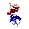
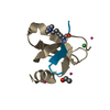
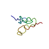
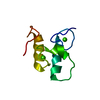
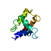
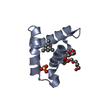
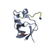
| Title | Beta-Scorpion Toxin folded in the periplasm of E.coli | ||||||
 Components Components | Beta-insect excitatory toxin Bj-xtrIT | ||||||
 Keywords Keywords | TOXIN / Alpha-Beta / Venom / Voltage Gated Na-Channels | ||||||
| Function / homology |  Function and homology information Function and homology informationsodium channel inhibitor activity / toxin activity / extracellular region Similarity search - Function | ||||||
| Biological species |  Hottentotta judaicus (scorpion) Hottentotta judaicus (scorpion) | ||||||
| Method |  X-RAY DIFFRACTION / SYNCHROTRON / MOLECULAR REPLACEMENT / Resolution: 1.7 Å X-RAY DIFFRACTION / SYNCHROTRON / MOLECULAR REPLACEMENT / Resolution: 1.7 Å | ||||||
 Authors Authors | O'Reilly, A.O. / Cole, A.R. / Lopes, J.L. / Lampert, A. / Wallace, B.A. | ||||||
 Citation Citation | Journal: Biochim.Biophys.Acta / Year: 2014 Title: Chaperone-mediated native folding of a beta-scorpion toxin in the periplasm of Escherichia coli. Authors: O'Reilly, A.O. / Cole, A.R. / Lopes, J.L. / Lampert, A. / Wallace, B.A. | ||||||
| History |
|
- Structure visualization
| Structure viewer | Molecule: MolmilJmol/JSmol |
|---|
- Downloads & links
-Download
| PDBx/mmCIF format | 4kyp.cif.gz | 77 KB | Display | PDBx/mmCIF format |
|---|---|---|---|---|
| PDB format | pdb4kyp.ent.gz | 58.2 KB | Display | PDB format |
| PDBx/mmJSON format | 4kyp.json.gz | Tree view | PDBx/mmJSON format | |
| Others |  Other downloads Other downloads |
-Validation report
| Arichive directory | https://data.pdbj.org/pub/pdb/validation_reports/ky/4kypftp://data.pdbj.org/pub/pdb/validation_reports/ky/4kyp | HTTPS FTP |
|---|
-Related structure data
| Related structure data | |
|---|---|
| Similar structure data |


-Links
 PDBj
PDBj

- Assembly
| Deposited unit | 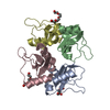
| |||||||||
|---|---|---|---|---|---|---|---|---|---|---|
| 1 | 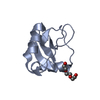
| |||||||||
| 2 | 
| |||||||||
| 3 | 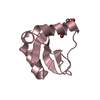
| |||||||||
| 4 | 
| |||||||||
| Unit cell |
| |||||||||
| Components on special symmetry positions |
|
-Components
| #1: Protein | Mass: 9439.647 Da / Num. of mol.: 4 Source method: isolated from a genetically manipulated source Source: (gene. exp.) Hottentotta judaicus (scorpion) / Gene: XTRIT / Production host:  #2: Chemical |   Mass: 150.173 Da / Num. of mol.: 2 / Source method: obtained synthetically / Formula: C6H14O4 Mass: 150.173 Da / Num. of mol.: 2 / Source method: obtained synthetically / Formula: C6H14O4#3: Chemical | ChemComp-PG4 / |   Mass: 194.226 Da / Num. of mol.: 1 / Source method: obtained synthetically / Formula: C8H18O5 / Comment: precipitant*YM Mass: 194.226 Da / Num. of mol.: 1 / Source method: obtained synthetically / Formula: C8H18O5 / Comment: precipitant*YM#4: Water | ChemComp-HOH / |  Mass: 18.015 Da / Num. of mol.: 339 / Source method: isolated from a natural source / Formula: H2O Mass: 18.015 Da / Num. of mol.: 339 / Source method: isolated from a natural source / Formula: H2OHas protein modification | Y | |
|---|
-Experimental details
-Experiment
| Experiment | Method: X-RAY DIFFRACTION / Number of used crystals: 1 |
|---|
- Sample preparation
| Crystal | Density Matthews: 2.25 Å3/Da / Density % sol: 45.42 % |
|---|---|
| Crystal grow | Temperature: 289 K / Method: vapor diffusion, hanging drop / pH: 5.5 Details: 0.2M NaCl, Bis-Tris, 29% PEG 3350 , pH 5.5, VAPOR DIFFUSION, HANGING DROP, temperature 289K |
-Data collection
| Diffraction | Mean temperature: 100 K |
|---|---|
| Diffraction source | Source: SYNCHROTRON / Site: SOLEIL  / Beamline: PROXIMA 1 / Wavelength: 0.98 Å / Beamline: PROXIMA 1 / Wavelength: 0.98 Å |
| Detector | Type: PSI PILATUS 6M / Detector: PIXEL / Date: Jun 23, 2012 |
| Radiation | Monochromator: channel cut cryogenically cooled monochromator crystal Protocol: SINGLE WAVELENGTH / Monochromatic (M) / Laue (L): M / Scattering type: x-ray |
| Radiation wavelength | Wavelength: 0.98 Å / Relative weight: 1 |
| Reflection | Resolution: 1.7→44.28 Å / Num. all: 26392 / Num. obs: 26392 / % possible obs: 69.23 % / Observed criterion σ(F): 1.48 / Observed criterion σ(I): 1.22 / Biso Wilson estimate: 20.05 Å2 |
| Reflection shell | Resolution: 1.7→1.74 Å / Redundancy: 1.2 % / Rmerge(I) obs: 0.709 / Mean I/σ(I) obs: 1.22 / % possible all: 9.8 |
- Processing
| Software |
| ||||||||||||||||||||||||||||||||||||||||||||||||||||||||||||||||||||||||||||||||||||||||||||||||||||||||||||||||||
|---|---|---|---|---|---|---|---|---|---|---|---|---|---|---|---|---|---|---|---|---|---|---|---|---|---|---|---|---|---|---|---|---|---|---|---|---|---|---|---|---|---|---|---|---|---|---|---|---|---|---|---|---|---|---|---|---|---|---|---|---|---|---|---|---|---|---|---|---|---|---|---|---|---|---|---|---|---|---|---|---|---|---|---|---|---|---|---|---|---|---|---|---|---|---|---|---|---|---|---|---|---|---|---|---|---|---|---|---|---|---|---|---|---|---|---|
| Refinement | Method to determine structure: MOLECULAR REPLACEMENT / Resolution: 1.7→44.28 Å / Cor.coef. Fo:Fc: 0.9112 / Cor.coef. Fo:Fc free: 0.8947 / Cross valid method: THROUGHOUT / σ(F): 0 / Stereochemistry target values: Engh & Huber
| ||||||||||||||||||||||||||||||||||||||||||||||||||||||||||||||||||||||||||||||||||||||||||||||||||||||||||||||||||
| Displacement parameters | Biso mean: 24.28 Å2
| ||||||||||||||||||||||||||||||||||||||||||||||||||||||||||||||||||||||||||||||||||||||||||||||||||||||||||||||||||
| Refine analyze | Luzzati coordinate error obs: 0.199 Å | ||||||||||||||||||||||||||||||||||||||||||||||||||||||||||||||||||||||||||||||||||||||||||||||||||||||||||||||||||
| Refinement step | Cycle: LAST / Resolution: 1.7→44.28 Å
| ||||||||||||||||||||||||||||||||||||||||||||||||||||||||||||||||||||||||||||||||||||||||||||||||||||||||||||||||||
| Refine LS restraints |
| ||||||||||||||||||||||||||||||||||||||||||||||||||||||||||||||||||||||||||||||||||||||||||||||||||||||||||||||||||
| LS refinement shell | Resolution: 1.7→1.77 Å / Total num. of bins used: 13
|
