 Movie
Movie Controller

[English] 日本語
 Yorodumi
- PDB-4icu: Ubiquitin-like domain of human tubulin folding cofactor E - cryst... -
+ Open data

- Basic information
| Entry | Database: PDB / ID: 4icu | ||||||
|---|---|---|---|---|---|---|---|
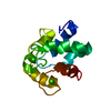
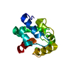
| Title | Ubiquitin-like domain of human tubulin folding cofactor E - crystal from A | ||||||
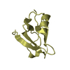
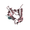
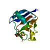
 Components Components | Tubulin-specific chaperone E | ||||||
 Keywords Keywords | CHAPERONE / Ubiquitin-like domain / tubulin folding cofactor / alpha tubulin / tubulin folding cofactor B | ||||||
| Function / homology |  Function and homology information Function and homology informationperipheral nervous system neuron axonogenesis / post-chaperonin tubulin folding pathway / muscle atrophy / Post-chaperonin tubulin folding pathway / tubulin complex assembly / developmental growth / alpha-tubulin binding / mitotic spindle organization / post-embryonic development / adult locomotory behavior ...peripheral nervous system neuron axonogenesis / post-chaperonin tubulin folding pathway / muscle atrophy / Post-chaperonin tubulin folding pathway / tubulin complex assembly / developmental growth / alpha-tubulin binding / mitotic spindle organization / post-embryonic development / adult locomotory behavior / microtubule cytoskeleton organization / protein-folding chaperone binding / protein folding / microtubule / cytoplasm Similarity search - Function | ||||||
| Biological species |  Homo sapiens (human) Homo sapiens (human) | ||||||
| Method |  X-RAY DIFFRACTION / SYNCHROTRON / MOLECULAR REPLACEMENT / Resolution: 2.4 Å X-RAY DIFFRACTION / SYNCHROTRON / MOLECULAR REPLACEMENT / Resolution: 2.4 Å | ||||||
 Authors Authors | Janowski, R. / Boutin, M. / Zabala, J.C. / Coll, M. | ||||||
 Citation Citation | Journal: J Cell Sci / Year: 2015 Title: The structure of the complex between α-tubulin, TBCE and TBCB reveals a tubulin dimer dissociation mechanism. Authors: Marina Serna / Gerardo Carranza / Jaime Martín-Benito / Robert Janowski / Albert Canals / Miquel Coll / Juan Carlos Zabala / José María Valpuesta /  Abstract: Tubulin proteostasis is regulated by a group of molecular chaperones termed tubulin cofactors (TBC). Whereas tubulin heterodimer formation is well-characterized biochemically, its dissociation ...Tubulin proteostasis is regulated by a group of molecular chaperones termed tubulin cofactors (TBC). Whereas tubulin heterodimer formation is well-characterized biochemically, its dissociation pathway is not clearly understood. Here, we carried out biochemical assays to dissect the role of the human TBCE and TBCB chaperones in α-tubulin-β-tubulin dissociation. We used electron microscopy and image processing to determine the three-dimensional structure of the human TBCE, TBCB and α-tubulin (αEB) complex, which is formed upon α-tubulin-β-tubulin heterodimer dissociation by the two chaperones. Docking the atomic structures of domains of these proteins, including the TBCE UBL domain, as we determined by X-ray crystallography, allowed description of the molecular architecture of the αEB complex. We found that heterodimer dissociation is an energy-independent process that takes place through a disruption of the α-tubulin-β-tubulin interface that is caused by a steric interaction between β-tubulin and the TBCE cytoskeleton-associated protein glycine-rich (CAP-Gly) and leucine-rich repeat (LRR) domains. The protruding arrangement of chaperone ubiquitin-like (UBL) domains in the αEB complex suggests that there is a direct interaction of this complex with the proteasome, thus mediating α-tubulin degradation. | ||||||
| History |
|
- Structure visualization
| Structure viewer | Molecule: MolmilJmol/JSmol |
|---|
- Downloads & links
-Download
| PDBx/mmCIF format | 4icu.cif.gz | 146.5 KB | Display | PDBx/mmCIF format |
|---|---|---|---|---|
| PDB format | pdb4icu.ent.gz | 118.3 KB | Display | PDB format |
| PDBx/mmJSON format | 4icu.json.gz | Tree view | PDBx/mmJSON format | |
| Others |  Other downloads Other downloads |
-Validation report
| Arichive directory | https://data.pdbj.org/pub/pdb/validation_reports/ic/4icuftp://data.pdbj.org/pub/pdb/validation_reports/ic/4icu | HTTPS FTP |
|---|
-Related structure data
| Related structure data |  2447C 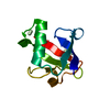 4icvSC S: Starting model for refinement C: citing same article ( |
|---|---|
| Similar structure data |


-Links
 PDBj
PDBj
- Assembly
| Deposited unit | 
| ||||||||||||||||||||||||||||||
|---|---|---|---|---|---|---|---|---|---|---|---|---|---|---|---|---|---|---|---|---|---|---|---|---|---|---|---|---|---|---|---|
| 1 | 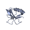
| ||||||||||||||||||||||||||||||
| 2 | 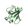
| ||||||||||||||||||||||||||||||
| 3 | 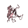
| ||||||||||||||||||||||||||||||
| 4 | 
| ||||||||||||||||||||||||||||||
| Unit cell |
| ||||||||||||||||||||||||||||||
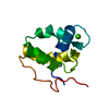
| Noncrystallographic symmetry (NCS) | NCS domain:
NCS domain segments: Component-ID: 1 / Ens-ID: 1 / Beg auth comp-ID: GLN / Beg label comp-ID: GLN / End auth comp-ID: TRP / End label comp-ID: TRP / Refine code: 2 / Auth seq-ID: 444 - 527 / Label seq-ID: 2 - 85
|
-Components
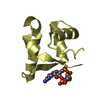
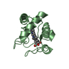
| #1: Protein | Mass: 9819.529 Da / Num. of mol.: 4 / Fragment: Ubiquitin-like domain, UNP residues 443-527 Source method: isolated from a genetically manipulated source Source: (gene. exp.) Homo sapiens (human) / Gene: TBCE / Production host:  #2: Water | ChemComp-HOH / |  Mass: 18.015 Da / Num. of mol.: 77 / Source method: isolated from a natural source / Formula: H2O Mass: 18.015 Da / Num. of mol.: 77 / Source method: isolated from a natural source / Formula: H2O |
|---|
-Experimental details
-Experiment
| Experiment | Method: X-RAY DIFFRACTION / Number of used crystals: 1 |
|---|
- Sample preparation
| Crystal | Density Matthews: 2.16 Å3/Da / Density % sol: 42.95 % |
|---|---|
| Crystal grow | Temperature: 292 K / Method: vapor diffusion, hanging drop / pH: 8.5 Details: 100 mM Tris pH 8.5, 200 mM sodium acetate and 26% PEG 4000, VAPOR DIFFUSION, HANGING DROP, temperature 292K |
-Data collection
| Diffraction | Mean temperature: 100 K |
|---|---|
| Diffraction source | Source: SYNCHROTRON / Site: ESRF  / Beamline: ID23-2 / Wavelength: 0.8726 Å / Beamline: ID23-2 / Wavelength: 0.8726 Å |
| Detector | Type: MARMOSAIC 225 mm CCD / Detector: CCD / Date: Feb 2, 2011 |
| Radiation | Protocol: SINGLE WAVELENGTH / Monochromatic (M) / Laue (L): M / Scattering type: x-ray |
| Radiation wavelength | Wavelength: 0.8726 Å / Relative weight: 1 |
| Reflection | Resolution: 2.4→30 Å / Num. obs: 13724 / % possible obs: 98.8 % / Observed criterion σ(F): 0 / Observed criterion σ(I): 0 / Redundancy: 6.9 % / Biso Wilson estimate: 23.53 Å2 / Rmerge(I) obs: 0.143 / Net I/σ(I): 11.2 |
| Reflection shell | Resolution: 2.4→2.46 Å / Rmerge(I) obs: 0.766 / Mean I/σ(I) obs: 2.3 / % possible all: 87.5 |
- Processing
| Software |
| |||||||||||||||||||||||||||||||||||||||||||||||||||||||||||||||||||||||||||||||||||||||||||||||||||||||||||||||||||||||||||||
|---|---|---|---|---|---|---|---|---|---|---|---|---|---|---|---|---|---|---|---|---|---|---|---|---|---|---|---|---|---|---|---|---|---|---|---|---|---|---|---|---|---|---|---|---|---|---|---|---|---|---|---|---|---|---|---|---|---|---|---|---|---|---|---|---|---|---|---|---|---|---|---|---|---|---|---|---|---|---|---|---|---|---|---|---|---|---|---|---|---|---|---|---|---|---|---|---|---|---|---|---|---|---|---|---|---|---|---|---|---|---|---|---|---|---|---|---|---|---|---|---|---|---|---|---|---|---|
| Refinement | Method to determine structure: MOLECULAR REPLACEMENT Starting model: PDB ENTRY 4ICV Resolution: 2.4→30 Å / Cor.coef. Fo:Fc: 0.942 / Cor.coef. Fo:Fc free: 0.908 / SU B: 20.031 / SU ML: 0.212 / Cross valid method: THROUGHOUT / σ(F): 0 / σ(I): 0 / ESU R: 0.649 / ESU R Free: 0.301 / Stereochemistry target values: MAXIMUM LIKELIHOOD
| |||||||||||||||||||||||||||||||||||||||||||||||||||||||||||||||||||||||||||||||||||||||||||||||||||||||||||||||||||||||||||||
| Solvent computation | Ion probe radii: 0.8 Å / Shrinkage radii: 0.8 Å / VDW probe radii: 1.4 Å / Solvent model: BABINET MODEL WITH MASK | |||||||||||||||||||||||||||||||||||||||||||||||||||||||||||||||||||||||||||||||||||||||||||||||||||||||||||||||||||||||||||||
| Displacement parameters | Biso mean: 39.579 Å2
| |||||||||||||||||||||||||||||||||||||||||||||||||||||||||||||||||||||||||||||||||||||||||||||||||||||||||||||||||||||||||||||
| Refinement step | Cycle: LAST / Resolution: 2.4→30 Å
| |||||||||||||||||||||||||||||||||||||||||||||||||||||||||||||||||||||||||||||||||||||||||||||||||||||||||||||||||||||||||||||
| Refine LS restraints |
| |||||||||||||||||||||||||||||||||||||||||||||||||||||||||||||||||||||||||||||||||||||||||||||||||||||||||||||||||||||||||||||
| Refine LS restraints NCS | Dom-ID: 1 / Ens-ID: 1 / Refine-ID: X-RAY DIFFRACTION
| |||||||||||||||||||||||||||||||||||||||||||||||||||||||||||||||||||||||||||||||||||||||||||||||||||||||||||||||||||||||||||||
| LS refinement shell | Resolution: 2.4→2.529 Å / Total num. of bins used: 10
| |||||||||||||||||||||||||||||||||||||||||||||||||||||||||||||||||||||||||||||||||||||||||||||||||||||||||||||||||||||||||||||
| Refinement TLS params. | Method: refined / Refine-ID: X-RAY DIFFRACTION
| |||||||||||||||||||||||||||||||||||||||||||||||||||||||||||||||||||||||||||||||||||||||||||||||||||||||||||||||||||||||||||||
| Refinement TLS group |
|
