 Movie
Movie Controller
 Structure viewers
 About Yorodumi Papers


+Search query
-Structure paper
| Title | The structure of the complex between α-tubulin, TBCE and TBCB reveals a tubulin dimer dissociation mechanism. |
|---|---|
| Journal, issue, pages | J Cell Sci, Vol. 128, Issue 9, Page 1824-1834, Year 2015 |
| Publish date | May 1, 2015 |
 Authors Authors | Marina Serna / Gerardo Carranza / Jaime Martín-Benito / Robert Janowski / Albert Canals / Miquel Coll / Juan Carlos Zabala / José María Valpuesta /  |
| PubMed Abstract | Tubulin proteostasis is regulated by a group of molecular chaperones termed tubulin cofactors (TBC). Whereas tubulin heterodimer formation is well-characterized biochemically, its dissociation ...Tubulin proteostasis is regulated by a group of molecular chaperones termed tubulin cofactors (TBC). Whereas tubulin heterodimer formation is well-characterized biochemically, its dissociation pathway is not clearly understood. Here, we carried out biochemical assays to dissect the role of the human TBCE and TBCB chaperones in α-tubulin-β-tubulin dissociation. We used electron microscopy and image processing to determine the three-dimensional structure of the human TBCE, TBCB and α-tubulin (αEB) complex, which is formed upon α-tubulin-β-tubulin heterodimer dissociation by the two chaperones. Docking the atomic structures of domains of these proteins, including the TBCE UBL domain, as we determined by X-ray crystallography, allowed description of the molecular architecture of the αEB complex. We found that heterodimer dissociation is an energy-independent process that takes place through a disruption of the α-tubulin-β-tubulin interface that is caused by a steric interaction between β-tubulin and the TBCE cytoskeleton-associated protein glycine-rich (CAP-Gly) and leucine-rich repeat (LRR) domains. The protruding arrangement of chaperone ubiquitin-like (UBL) domains in the αEB complex suggests that there is a direct interaction of this complex with the proteasome, thus mediating α-tubulin degradation. |
 External links External links | J Cell Sci / PubMed:25908846 |
| Methods | EM (single particle) / X-ray diffraction |
| Resolution | 1.45 - 21.0 Å |
| Structure data | 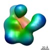 EMDB-2447: 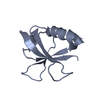 PDB-4icu: 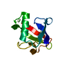 PDB-4icv: |
| Chemicals |  ChemComp-HOH:  ChemComp-PR: |
| Source |
|
 Keywords Keywords | CHAPERONE / Ubiquitin-like domain / tubulin folding cofactor / alpha tubulin / tubulin folding cofactor B |
 homo sapiens (human)
