 Movie
Movie Controller

[English] 日本語
 Yorodumi
- PDB-1iqy: CRYSTAL STRUCTURE OF NICKEL-SUBSTITUTED AMINE OXIDASE FROM ARTHRO... -
+ Open data

- Basic information
| Entry | Database: PDB / ID: 1iqy | ||||||
|---|---|---|---|---|---|---|---|
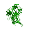
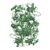
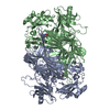
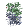
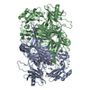
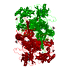
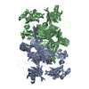
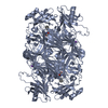
| Title | CRYSTAL STRUCTURE OF NICKEL-SUBSTITUTED AMINE OXIDASE FROM ARTHROBACTER GLOBIFORMIS | ||||||
 Components Components | AMINE OXIDASE | ||||||
 Keywords Keywords | OXIDOREDUCTASE / COPPER / AMINE OXIDASE / ARTHROBACTER GLOBIFORMIS / QUINONE COFACTOR / TPQ / NICKEL / NI(II) | ||||||
| Function / homology |  Function and homology information Function and homology informationprimary-amine oxidase / primary methylamine oxidase activity / amine metabolic process / quinone binding / copper ion binding Similarity search - Function | ||||||
| Biological species |  Arthrobacter globiformis (bacteria) Arthrobacter globiformis (bacteria) | ||||||
| Method |  X-RAY DIFFRACTION / SYNCHROTRON / MOLECULAR REPLACEMENT / Resolution: 1.8 Å X-RAY DIFFRACTION / SYNCHROTRON / MOLECULAR REPLACEMENT / Resolution: 1.8 Å | ||||||
 Authors Authors | Kishishita, S. / Okajima, T. / Mure, M. / Kim, M. / Yamaguchi, H. / Hirota, S. / Kuroda, S. / Tanizawa, K. | ||||||
 Citation Citation | Journal: J.AM.CHEM.SOC. / Year: 2003 Title: Role of Copper Ion in Bacterial Copper Amine Oxidase: Spectroscopic and Crystallographic Studies of Metal-Substituted Enzymes Authors: Kishishita, S. / Okajima, T. / Kim, M. / Yamaguchi, H. / Hirota, S. / Suzuki, S. / Kuroda, S. / Tanizawa, K. / Mure, M. | ||||||
| History |
|
- Structure visualization
| Structure viewer | Molecule: MolmilJmol/JSmol |
|---|
- Downloads & links
-Download
| PDBx/mmCIF format | 1iqy.cif.gz | 271.2 KB | Display | PDBx/mmCIF format |
|---|---|---|---|---|
| PDB format | pdb1iqy.ent.gz | 216.8 KB | Display | PDB format |
| PDBx/mmJSON format | 1iqy.json.gz | Tree view | PDBx/mmJSON format | |
| Others |  Other downloads Other downloads |
-Validation report
| Arichive directory | https://data.pdbj.org/pub/pdb/validation_reports/iq/1iqyftp://data.pdbj.org/pub/pdb/validation_reports/iq/1iqy | HTTPS FTP |
|---|
-Related structure data


-Links
 PDBj
PDBj- Assembly
| Deposited unit | 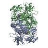
| ||||||||
|---|---|---|---|---|---|---|---|---|---|
| 1 |
| ||||||||
| Unit cell |
|
-Components
| #1: Protein | Mass: 70752.742 Da / Num. of mol.: 2 Source method: isolated from a genetically manipulated source Source: (gene. exp.) Arthrobacter globiformis (bacteria) / Plasmid: PEPO2 / Species (production host): Escherichia coli / Production host: #2: Chemical |   Mass: 58.693 Da / Num. of mol.: 2 / Source method: obtained synthetically / Formula: Ni Mass: 58.693 Da / Num. of mol.: 2 / Source method: obtained synthetically / Formula: Ni#3: Water | ChemComp-HOH / |  Mass: 18.015 Da / Num. of mol.: 908 / Source method: isolated from a natural source / Formula: H2O Mass: 18.015 Da / Num. of mol.: 908 / Source method: isolated from a natural source / Formula: H2O |
|---|
-Experimental details
-Experiment
| Experiment | Method: X-RAY DIFFRACTION / Number of used crystals: 1 |
|---|
- Sample preparation
| Crystal | Density Matthews: 2.99 Å3/Da / Density % sol: 58.9 % | ||||||||||||||||||||||||
|---|---|---|---|---|---|---|---|---|---|---|---|---|---|---|---|---|---|---|---|---|---|---|---|---|---|
| Crystal grow | Temperature: 288 K / Method: microdialysis / pH: 6.8 Details: potassium-sodium tartrate, pH 6.8, MICRODIALYSIS, temperature 288K | ||||||||||||||||||||||||
| Crystal grow | *PLUS Temperature: 16 ℃ | ||||||||||||||||||||||||
| Components of the solutions | *PLUS
|
-Data collection
| Diffraction | Mean temperature: 100 K |
|---|---|
| Diffraction source | Source: SYNCHROTRON / Site: Photon Factory  / Beamline: BL-18B / Wavelength: 1 / Wavelength: 1 Å / Beamline: BL-18B / Wavelength: 1 / Wavelength: 1 Å |
| Detector | Type: RIGAKU RAXIS IV / Detector: IMAGE PLATE / Date: Jun 24, 1999 |
| Radiation | Monochromator: Si 111 / Protocol: SINGLE WAVELENGTH / Monochromatic (M) / Laue (L): M / Scattering type: x-ray |
| Radiation wavelength | Wavelength: 1 Å / Relative weight: 1 |
| Reflection | Resolution: 1.8→10 Å / Num. all: 155164 / Num. obs: 146419 / % possible obs: 94.8 % / Observed criterion σ(F): 1 / Observed criterion σ(I): 3 / Rmerge(I) obs: 0.038 |
| Reflection shell | Resolution: 1.8→1.86 Å / % possible all: 81.4 |
| Reflection | *PLUS Lowest resolution: 10 Å / Num. measured all: 364483 |
- Processing
| Software |
| |||||||||||||||||||||||||
|---|---|---|---|---|---|---|---|---|---|---|---|---|---|---|---|---|---|---|---|---|---|---|---|---|---|---|
| Refinement | Method to determine structure: MOLECULAR REPLACEMENT Starting model: HOLO-AGAO Resolution: 1.8→10 Å / Data cutoff high absF: 1000000 / Data cutoff low absF: 0.001 / Cross valid method: THROUGHOUT / σ(F): 2 / σ(I): 0 / Stereochemistry target values: Engh & Huber
| |||||||||||||||||||||||||
| Displacement parameters | Biso mean: 21.8 Å2 | |||||||||||||||||||||||||
| Refinement step | Cycle: LAST / Resolution: 1.8→10 Å
| |||||||||||||||||||||||||
| Refine LS restraints |
| |||||||||||||||||||||||||
| LS refinement shell | Resolution: 1.8→1.88 Å
| |||||||||||||||||||||||||
| Xplor file |
| |||||||||||||||||||||||||
| Refinement | *PLUS Lowest resolution: 10 Å / % reflection Rfree: 5 % / Rfactor Rfree: 0.26 | |||||||||||||||||||||||||
| Solvent computation | *PLUS | |||||||||||||||||||||||||
| Displacement parameters | *PLUS | |||||||||||||||||||||||||
| Refine LS restraints | *PLUS
|
