 Movie
Movie Controller


+ Open data

- Basic information
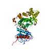
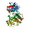
| Entry | Database: PDB / ID: 1imv | ||||||
|---|---|---|---|---|---|---|---|
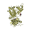
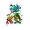
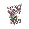
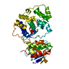
| Title | 2.85 A crystal structure of PEDF | ||||||
 Components Components | PIGMENT EPITHELIUM-DERIVED FACTOR | ||||||
 Keywords Keywords | SIGNALING PROTEIN / serpin / PEDF / angiogenesis | ||||||
| Function / homology |  Function and homology information Function and homology informationcellular response to cobalt ion / ovulation cycle / response to peptide / negative regulation of epithelial cell proliferation involved in prostate gland development / axon hillock / epithelial cell proliferation involved in prostate gland development / response to arsenic-containing substance / short-term memory / negative regulation of endothelial cell migration / response to acidic pH ...cellular response to cobalt ion / ovulation cycle / response to peptide / negative regulation of epithelial cell proliferation involved in prostate gland development / axon hillock / epithelial cell proliferation involved in prostate gland development / response to arsenic-containing substance / short-term memory / negative regulation of endothelial cell migration / response to acidic pH / positive regulation of neurogenesis / basement membrane / cellular response to dexamethasone stimulus / cellular response to retinoic acid / negative regulation of angiogenesis / kidney development / serine-type endopeptidase inhibitor activity / cellular response to glucose stimulus / positive regulation of neuron projection development / : / melanosome / retina development in camera-type eye / negative regulation of gene expression / perinuclear region of cytoplasm / extracellular space / extracellular exosome / extracellular region Similarity search - Function | ||||||
| Biological species |  Homo sapiens (human) Homo sapiens (human) | ||||||
| Method |  X-RAY DIFFRACTION / MOLECULAR REPLACEMENT / Resolution: 2.85 Å X-RAY DIFFRACTION / MOLECULAR REPLACEMENT / Resolution: 2.85 Å | ||||||
 Authors Authors | Simonovic, M. / Gettins, P.G.W. / Volz, K. | ||||||
 Citation Citation | Journal: Proc.Natl.Acad.Sci.USA / Year: 2001 Title: Crystal structure of human PEDF, a potent anti-angiogenic and neurite growth-promoting factor. Authors: Simonovic, M. / Gettins, P.G. / Volz, K. | ||||||
| History |
|
- Structure visualization
| Structure viewer | Molecule: MolmilJmol/JSmol |
|---|
- Downloads & links
-Download
| PDBx/mmCIF format | 1imv.cif.gz | 86.7 KB | Display | PDBx/mmCIF format |
|---|---|---|---|---|
| PDB format | pdb1imv.ent.gz | 64.9 KB | Display | PDB format |
| PDBx/mmJSON format | 1imv.json.gz | Tree view | PDBx/mmJSON format | |
| Others |  Other downloads Other downloads |
-Validation report
| Arichive directory | https://data.pdbj.org/pub/pdb/validation_reports/im/1imvftp://data.pdbj.org/pub/pdb/validation_reports/im/1imv | HTTPS FTP |
|---|
-Related structure data
| Related structure data |  1qlpS S: Starting model for refinement |
|---|---|
| Similar structure data |
-Links
 PDBj
PDBj
- Assembly
| Deposited unit | 
| ||||||||
|---|---|---|---|---|---|---|---|---|---|
| 1 |
| ||||||||
| Unit cell |
|
-Components
| #1: Protein | Mass: 44328.516 Da / Num. of mol.: 1 Source method: isolated from a genetically manipulated source Source: (gene. exp.) Homo sapiens (human) / Plasmid: pMA / Cell line (production host): BHK / Production host:   Cricetulus griseus (Chinese hamster) / References: UniProt: P36955 Cricetulus griseus (Chinese hamster) / References: UniProt: P36955 |
|---|---|
| #2: Sugar | ChemComp-NAG /   Type: D-saccharide, beta linking / Mass: 221.208 Da / Num. of mol.: 1 Type: D-saccharide, beta linking / Mass: 221.208 Da / Num. of mol.: 1Source method: isolated from a genetically manipulated source Formula: C8H15NO6 |
| #3: Water | ChemComp-HOH /  Mass: 18.015 Da / Num. of mol.: 45 / Source method: isolated from a natural source / Formula: H2O Mass: 18.015 Da / Num. of mol.: 45 / Source method: isolated from a natural source / Formula: H2O |
| Has protein modification | Y |
-Experimental details
-Experiment
| Experiment | Method: X-RAY DIFFRACTION / Number of used crystals: 3 |
|---|
- Sample preparation
| Crystal | Density Matthews: 2.82 Å3/Da / Density % sol: 56.36 % | ||||||||||||||||||||
|---|---|---|---|---|---|---|---|---|---|---|---|---|---|---|---|---|---|---|---|---|---|
| Crystal grow | Temperature: 293 K / Method: vapor diffusion, hanging drop / pH: 6.2 Details: 0.2M ammonium fluoride, 20% PEG 3350, pH 6.20, VAPOR DIFFUSION, HANGING DROP, temperature 293K | ||||||||||||||||||||
| Crystal | *PLUS Density % sol: 58 % | ||||||||||||||||||||
| Crystal grow | *PLUS Temperature: 18 ℃ | ||||||||||||||||||||
| Components of the solutions | *PLUS
|
-Data collection
| Diffraction |
| ||||||||||||||||||||||||
|---|---|---|---|---|---|---|---|---|---|---|---|---|---|---|---|---|---|---|---|---|---|---|---|---|---|
| Diffraction source |
| ||||||||||||||||||||||||
| Detector |
| ||||||||||||||||||||||||
| Radiation |
| ||||||||||||||||||||||||
| Radiation wavelength | Wavelength: 1.5418 Å / Relative weight: 1 | ||||||||||||||||||||||||
| Reflection | Resolution: 2.85→36.74 Å / Num. all: 11214 / Num. obs: 11214 / % possible obs: 92.1 % / Observed criterion σ(F): 0 / Observed criterion σ(I): 0 / Redundancy: 12.7 % / Biso Wilson estimate: 45.2 Å2 / Rmerge(I) obs: 0.127 / Net I/σ(I): 8.5 | ||||||||||||||||||||||||
| Reflection shell | Resolution: 2.85→3.03 Å / Redundancy: 3 % / Rmerge(I) obs: 0.37 / Num. unique all: 1166 / % possible all: 65.4 | ||||||||||||||||||||||||
| Reflection | *PLUS Num. measured all: 152138 | ||||||||||||||||||||||||
| Reflection shell | *PLUS % possible obs: 65.4 % |
- Processing
| Software |
| ||||||||||||||||||||||||||||||||||||||||
|---|---|---|---|---|---|---|---|---|---|---|---|---|---|---|---|---|---|---|---|---|---|---|---|---|---|---|---|---|---|---|---|---|---|---|---|---|---|---|---|---|---|
| Refinement | Method to determine structure: MOLECULAR REPLACEMENT Starting model: PDB entry 1QLP Resolution: 2.85→36.74 Å / Rfactor Rfree error: 0.007 / Data cutoff high absF: 200608.11 / Data cutoff low absF: 0 / Isotropic thermal model: RESTRAINED / Cross valid method: THROUGHOUT / σ(F): 0 / σ(I): 0 / Stereochemistry target values: Engh & Huber Details: Residues 1-15 and 353-360 are missing/disordered. The side chains of the following residues are disordered: THR16, ARG79, LYS126, LYS127, ARG174, LYS177, GLU178, ASP181, GLU182, LYS228, THR352.
| ||||||||||||||||||||||||||||||||||||||||
| Solvent computation | Solvent model: FLAT MODEL / Bsol: 36.78 Å2 / ksol: 0.32 e/Å3 | ||||||||||||||||||||||||||||||||||||||||
| Displacement parameters | Biso mean: 31.4 Å2
| ||||||||||||||||||||||||||||||||||||||||
| Refine analyze |
| ||||||||||||||||||||||||||||||||||||||||
| Refinement step | Cycle: LAST / Resolution: 2.85→36.74 Å
| ||||||||||||||||||||||||||||||||||||||||
| Refine LS restraints |
| ||||||||||||||||||||||||||||||||||||||||
| LS refinement shell | Resolution: 2.85→3.03 Å / Rfactor Rfree error: 0.029 / Total num. of bins used: 6
| ||||||||||||||||||||||||||||||||||||||||
| Xplor file |
| ||||||||||||||||||||||||||||||||||||||||
| Software | *PLUS Name: CNS / Version: 1 / Classification: refinement | ||||||||||||||||||||||||||||||||||||||||
| Refinement | *PLUS σ(F): 0 / % reflection Rfree: 10.4 % / Rfactor obs: 0.188 | ||||||||||||||||||||||||||||||||||||||||
| Solvent computation | *PLUS | ||||||||||||||||||||||||||||||||||||||||
| Displacement parameters | *PLUS Biso mean: 31.4 Å2 | ||||||||||||||||||||||||||||||||||||||||
| Refine LS restraints | *PLUS
| ||||||||||||||||||||||||||||||||||||||||
| LS refinement shell | *PLUS Rfactor Rfree: 0.34 / % reflection Rfree: 10.7 % / Rfactor Rwork: 0.301 |
