 Movie
Movie Controller

[English] 日本語
 Yorodumi
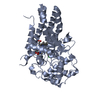
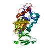
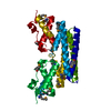
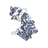
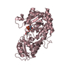
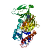
- PDB-3t9c: Crystal structure of the catalytic domain of human diphosphoinosi... -
+ Open data

- Basic information
| Entry | Database: PDB / ID: 3t9c | ||||||
|---|---|---|---|---|---|---|---|
| Title | Crystal structure of the catalytic domain of human diphosphoinositol pentakisphosphate kinase 2 (PPIP5K2) in complex with AMPPNP and inositol hexakisphosphate (IP6) | ||||||
 Components Components | Inositol Pyrophosphate Kinase | ||||||
 Keywords Keywords | TRANSFERASE / ATP-grasp Fold / Inositol Pyrophosphate Kinase / Phosphoryl Transferase | ||||||
| Function / homology |  Function and homology information Function and homology informationinositol-1,3,4,5,6-pentakisphosphate kinase activity / diphosphoinositol pentakisphosphate kinase activity / inositol hexakisphosphate 5-kinase activity / diphosphoinositol-pentakisphosphate 1-kinase / 5-diphosphoinositol pentakisphosphate 1-kinase activity / inositol hexakisphosphate 1-kinase activity / inositol hexakisphosphate kinase activity / Synthesis of pyrophosphates in the cytosol / inositol phosphate metabolic process / inositol metabolic process ...inositol-1,3,4,5,6-pentakisphosphate kinase activity / diphosphoinositol pentakisphosphate kinase activity / inositol hexakisphosphate 5-kinase activity / diphosphoinositol-pentakisphosphate 1-kinase / 5-diphosphoinositol pentakisphosphate 1-kinase activity / inositol hexakisphosphate 1-kinase activity / inositol hexakisphosphate kinase activity / Synthesis of pyrophosphates in the cytosol / inositol phosphate metabolic process / inositol metabolic process / inositol phosphate biosynthetic process / sensory perception of sound / ATP binding / cytosol Similarity search - Function | ||||||
| Biological species |  Homo sapiens (human) Homo sapiens (human) | ||||||
| Method |  X-RAY DIFFRACTION / FOURIER SYNTHESIS / Resolution: 1.9 Å X-RAY DIFFRACTION / FOURIER SYNTHESIS / Resolution: 1.9 Å | ||||||
 Authors Authors | Wang, H. / Falck, J. / Hall, T.M.T. / Shears, S.B. | ||||||
 Citation Citation | Journal: Nat.Chem.Biol. / Year: 2011 Title: Structural basis for an inositol pyrophosphate kinase surmounting phosphate crowding. Authors: Wang, H. / Falck, J.R. / Hall, T.M. / Shears, S.B. | ||||||
| History |
|
- Structure visualization
| Structure viewer | Molecule: MolmilJmol/JSmol |
|---|
- Downloads & links
-Download
| PDBx/mmCIF format | 3t9c.cif.gz | 96.1 KB | Display | PDBx/mmCIF format |
|---|---|---|---|---|
| PDB format | pdb3t9c.ent.gz | 69.3 KB | Display | PDB format |
| PDBx/mmJSON format | 3t9c.json.gz | Tree view | PDBx/mmJSON format | |
| Others |  Other downloads Other downloads |
-Validation report
| Arichive directory | https://data.pdbj.org/pub/pdb/validation_reports/t9/3t9cftp://data.pdbj.org/pub/pdb/validation_reports/t9/3t9c | HTTPS FTP |
|---|
-Related structure data
| Related structure data |  3t54SC  3t7aC  3t99C  3t9aC  3t9bC  3t9dC  3t9eC 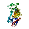 3t9fC S: Starting model for refinement C: citing same article ( |
|---|---|
| Similar structure data |


-Links
 PDBj
PDBj


- Assembly
| Deposited unit | 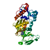
| ||||||||
|---|---|---|---|---|---|---|---|---|---|
| 1 |
| ||||||||
| Unit cell |
|
-Components
| #1: Protein | Mass: 37568.891 Da / Num. of mol.: 1 / Fragment: ATP-grasp Kinase domain Source method: isolated from a genetically manipulated source Source: (gene. exp.) Homo sapiens (human) / Gene: PPIP5K2 / Plasmid: pDest566 / Production host:  References: UniProt: O43314, diphosphoinositol-pentakisphosphate 1-kinase | ||
|---|---|---|---|
| #2: Chemical | ChemComp-ANP /   Mass: 506.196 Da / Num. of mol.: 1 / Source method: obtained synthetically / Formula: C10H17N6O12P3 / Comment: AMP-PNP, energy-carrying molecule analogue*YM Mass: 506.196 Da / Num. of mol.: 1 / Source method: obtained synthetically / Formula: C10H17N6O12P3 / Comment: AMP-PNP, energy-carrying molecule analogue*YM | ||
| #3: Chemical | ChemComp-IHP /   Mass: 660.035 Da / Num. of mol.: 1 / Source method: obtained synthetically / Formula: C6H18O24P6 Mass: 660.035 Da / Num. of mol.: 1 / Source method: obtained synthetically / Formula: C6H18O24P6 | ||
| #4: Chemical | ChemComp-MG /   Mass: 24.305 Da / Num. of mol.: 4 / Source method: obtained synthetically / Formula: Mg Mass: 24.305 Da / Num. of mol.: 4 / Source method: obtained synthetically / Formula: Mg#5: Water | ChemComp-HOH / |  Mass: 18.015 Da / Num. of mol.: 424 / Source method: isolated from a natural source / Formula: H2O Mass: 18.015 Da / Num. of mol.: 424 / Source method: isolated from a natural source / Formula: H2O |
-Experimental details
-Experiment
| Experiment | Method: X-RAY DIFFRACTION / Number of used crystals: 1 |
|---|
- Sample preparation
| Crystal | Density Matthews: 2.71 Å3/Da / Density % sol: 54.55 % |
|---|---|
| Crystal grow | Temperature: 277 K / Method: vapor diffusion, hanging drop Details: 12% (w/v) PEG 3350, 20 mM MgCl2, 0.1 M HEPES, pH 7.0, 1 mM AMPPNP, 2 mM CdCl2, Soaking with 10mM IP6 and 10 mM NaF under pH 5.2 for 3 days, VAPOR DIFFUSION, HANGING DROP, temperature 277K |
-Data collection
| Diffraction | Mean temperature: 100 K |
|---|---|
| Diffraction source | Source: ROTATING ANODE / Type: RIGAKU MICROMAX-007 HF / Wavelength: 1.5418 Å |
| Detector | Type: RIGAKU SATURN 92 / Detector: CCD / Date: Mar 5, 2011 |
| Radiation | Protocol: SINGLE WAVELENGTH / Monochromatic (M) / Laue (L): M / Scattering type: x-ray |
| Radiation wavelength | Wavelength: 1.5418 Å / Relative weight: 1 |
| Reflection | Resolution: 1.9→50 Å / Num. obs: 32957 / % possible obs: 99.7 % / Observed criterion σ(F): -3 / Observed criterion σ(I): 0 / Redundancy: 6.1 % / Rsym value: 0.062 / Net I/σ(I): 26.1 |
| Reflection shell | Resolution: 1.9→1.93 Å / Redundancy: 3.2 % / Mean I/σ(I) obs: 3.1 / Rsym value: 0.362 / % possible all: 77.4 |
- Processing
| Software |
| |||||||||||||||||||||||||||||||||||||||||||||||||||||||||||||||||
|---|---|---|---|---|---|---|---|---|---|---|---|---|---|---|---|---|---|---|---|---|---|---|---|---|---|---|---|---|---|---|---|---|---|---|---|---|---|---|---|---|---|---|---|---|---|---|---|---|---|---|---|---|---|---|---|---|---|---|---|---|---|---|---|---|---|---|
| Refinement | Method to determine structure: FOURIER SYNTHESIS Starting model: PDB ENTRY 3T54 Resolution: 1.9→23.55 Å / Cor.coef. Fo:Fc: 0.947 / Cor.coef. Fo:Fc free: 0.93 / Cross valid method: THROUGHOUT / σ(F): 0 / ESU R Free: 0.136 / Stereochemistry target values: MAXIMUM LIKELIHOOD
| |||||||||||||||||||||||||||||||||||||||||||||||||||||||||||||||||
| Solvent computation | Ion probe radii: 0.8 Å / Shrinkage radii: 0.8 Å / VDW probe radii: 1.4 Å / Solvent model: MASK | |||||||||||||||||||||||||||||||||||||||||||||||||||||||||||||||||
| Displacement parameters | Biso mean: 19.473 Å2
| |||||||||||||||||||||||||||||||||||||||||||||||||||||||||||||||||
| Refinement step | Cycle: LAST / Resolution: 1.9→23.55 Å
| |||||||||||||||||||||||||||||||||||||||||||||||||||||||||||||||||
| Refine LS restraints |
| |||||||||||||||||||||||||||||||||||||||||||||||||||||||||||||||||
| LS refinement shell | Resolution: 1.9→1.948 Å / Total num. of bins used: 20
|
