 Movie
Movie Controller

[English] 日本語
 Yorodumi
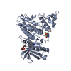
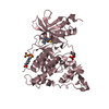
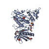
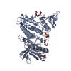
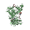
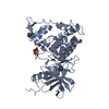
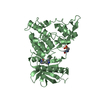
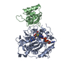
- PDB-4mq1: The crystal structure of DYRK1a with a bound pyrido[2,3-d]pyrimid... -
+ Open data

- Basic information
| Entry | Database: PDB / ID: 4mq1 | ||||||
|---|---|---|---|---|---|---|---|
| Title | The crystal structure of DYRK1a with a bound pyrido[2,3-d]pyrimidine inhibitor | ||||||
 Components Components | Dual specificity tyrosine-phosphorylation-regulated kinase 1A | ||||||
 Keywords Keywords | TRANSFERASE/TRANSFERASE INHIBITOR / dyrk1a / dyrk1b / kinase / TRANSFERASE-TRANSFERASE INHIBITOR complex | ||||||
| Function / homology |  Function and homology information Function and homology informationregulation of amyloid-beta formation / regulation of neurofibrillary tangle assembly / histone H3T45 kinase activity / negative regulation of heterochromatin formation / dual-specificity kinase / splicing factor binding / [RNA-polymerase]-subunit kinase / tau-protein kinase activity / regulation of alternative mRNA splicing, via spliceosome / negative regulation of microtubule polymerization ...regulation of amyloid-beta formation / regulation of neurofibrillary tangle assembly / histone H3T45 kinase activity / negative regulation of heterochromatin formation / dual-specificity kinase / splicing factor binding / [RNA-polymerase]-subunit kinase / tau-protein kinase activity / regulation of alternative mRNA splicing, via spliceosome / negative regulation of microtubule polymerization / negative regulation of DNA damage response, signal transduction by p53 class mediator / negative regulation of mRNA splicing, via spliceosome / G0 and Early G1 / cytoskeletal protein binding / peptidyl-tyrosine phosphorylation / RNA polymerase II CTD heptapeptide repeat kinase activity / protein serine/threonine/tyrosine kinase activity / positive regulation of RNA splicing / non-membrane spanning protein tyrosine kinase activity / tubulin binding / circadian rhythm / tau protein binding / protein autophosphorylation / nervous system development / actin binding / protein tyrosine kinase activity / protein phosphorylation / protein kinase activity / transcription coactivator activity / nuclear speck / ribonucleoprotein complex / protein serine kinase activity / axon / protein serine/threonine kinase activity / centrosome / dendrite / positive regulation of DNA-templated transcription / nucleoplasm / ATP binding / identical protein binding / nucleus / cytoplasm / cytosol Similarity search - Function | ||||||
| Biological species |  Homo sapiens (human) Homo sapiens (human) | ||||||
| Method |  X-RAY DIFFRACTION / SYNCHROTRON / MOLECULAR REPLACEMENT / Resolution: 2.35 Å X-RAY DIFFRACTION / SYNCHROTRON / MOLECULAR REPLACEMENT / Resolution: 2.35 Å | ||||||
 Authors Authors | Lukacs, C.M. / Janson, C.A. / Garvie, C. / Liang, L. | ||||||
 Citation Citation | Journal: Bioorg.Med.Chem.Lett. / Year: 2013 Title: Pyrido[2,3-d]pyrimidines: Discovery and preliminary SAR of a novel series of DYRK1B and DYRK1A inhibitors. Authors: Anderson, K. / Chen, Y. / Chen, Z. / Dominique, R. / Glenn, K. / He, Y. / Janson, C. / Luk, K.C. / Lukacs, C. / Polonskaia, A. / Qiao, Q. / Railkar, A. / Rossman, P. / Sun, H. / Xiang, Q. / ...Authors: Anderson, K. / Chen, Y. / Chen, Z. / Dominique, R. / Glenn, K. / He, Y. / Janson, C. / Luk, K.C. / Lukacs, C. / Polonskaia, A. / Qiao, Q. / Railkar, A. / Rossman, P. / Sun, H. / Xiang, Q. / Vilenchik, M. / Wovkulich, P. / Zhang, X. | ||||||
| History |
|
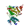
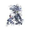
- Structure visualization
| Structure viewer | Molecule: MolmilJmol/JSmol |
|---|
- Downloads & links
-Download
| PDBx/mmCIF format | 4mq1.cif.gz | 298.9 KB | Display | PDBx/mmCIF format |
|---|---|---|---|---|
| PDB format | pdb4mq1.ent.gz | 242.7 KB | Display | PDB format |
| PDBx/mmJSON format | 4mq1.json.gz | Tree view | PDBx/mmJSON format | |
| Others |  Other downloads Other downloads |
-Validation report
| Arichive directory | https://data.pdbj.org/pub/pdb/validation_reports/mq/4mq1ftp://data.pdbj.org/pub/pdb/validation_reports/mq/4mq1 | HTTPS FTP |
|---|
-Related structure data
| Related structure data |  4mq2C 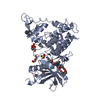 2vx3S S: Starting model for refinement C: citing same article ( |
|---|---|
| Similar structure data |

-Links
 PDBj
PDBj
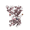
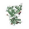
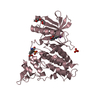
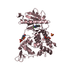
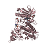
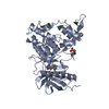
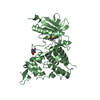
- Assembly
| Deposited unit | 
| ||||||||||||||||||||
|---|---|---|---|---|---|---|---|---|---|---|---|---|---|---|---|---|---|---|---|---|---|
| 1 | 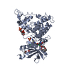
| ||||||||||||||||||||
| 2 | 
| ||||||||||||||||||||
| 3 | 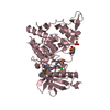
| ||||||||||||||||||||
| 4 | 
| ||||||||||||||||||||
| Unit cell |
| ||||||||||||||||||||
| Components on special symmetry positions |
| ||||||||||||||||||||
| Noncrystallographic symmetry (NCS) | NCS oper:
|
-Components
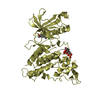
| #1: Protein | Mass: 42081.535 Da / Num. of mol.: 4 / Fragment: UNP residues 127-485 Source method: isolated from a genetically manipulated source Source: (gene. exp.) Homo sapiens (human) / Gene: DYRK1A, DYRK, MNB, MNBH / Plasmid: pNIC28-Bsa4 / Production host:  #2: Chemical | ChemComp-2C3 /   Mass: 541.386 Da / Num. of mol.: 4 / Source method: obtained synthetically / Formula: C25H22Cl2N6O4 Mass: 541.386 Da / Num. of mol.: 4 / Source method: obtained synthetically / Formula: C25H22Cl2N6O4#3: Chemical | ChemComp-1PE /   Mass: 238.278 Da / Num. of mol.: 6 / Source method: obtained synthetically / Formula: C10H22O6 / Comment: precipitant*YM Mass: 238.278 Da / Num. of mol.: 6 / Source method: obtained synthetically / Formula: C10H22O6 / Comment: precipitant*YM#4: Chemical | ChemComp-SO4 /   Mass: 96.063 Da / Num. of mol.: 8 / Source method: obtained synthetically / Formula: SO4 Mass: 96.063 Da / Num. of mol.: 8 / Source method: obtained synthetically / Formula: SO4#5: Water | ChemComp-HOH / |  Mass: 18.015 Da / Num. of mol.: 470 / Source method: isolated from a natural source / Formula: H2O Mass: 18.015 Da / Num. of mol.: 470 / Source method: isolated from a natural source / Formula: H2OHas protein modification | Y | |
|---|
-Experimental details
-Experiment
| Experiment | Method: X-RAY DIFFRACTION / Number of used crystals: 1 |
|---|
- Sample preparation
| Crystal | Density Matthews: 3.24 Å3/Da / Density % sol: 61.98 % |
|---|---|
| Crystal grow | Temperature: 277 K / Method: vapor diffusion, sitting drop / pH: 8.5 Details: 15% PEG 300, 0.1M LiSO4, 0.1M Tris, pH 8.5, VAPOR DIFFUSION, SITTING DROP, temperature 277K |
-Data collection
| Diffraction | Mean temperature: 100 K |
|---|---|
| Diffraction source | Source: SYNCHROTRON / Site: APS  / Beamline: 31-ID / Wavelength: 0.97929 Å / Beamline: 31-ID / Wavelength: 0.97929 Å |
| Detector | Type: RAYONIX MX225HE / Detector: CCD / Date: Dec 3, 2012 |
| Radiation | Protocol: SINGLE WAVELENGTH / Monochromatic (M) / Laue (L): M / Scattering type: x-ray |
| Radiation wavelength | Wavelength: 0.97929 Å / Relative weight: 1 |
| Reflection | Resolution: 2.35→50 Å / Num. obs: 90023 / % possible obs: 99.4 % / Redundancy: 3.8 % / Biso Wilson estimate: 53 Å2 / Rmerge(I) obs: 0.09 / Net I/σ(I): 8.9 |
- Processing
| Software |
| ||||||||||||||||||||||||||||||||||||||||||||||||||||||||||||||||||||||||||||||||||||||||||||||||||||||||||||||||||||||||||||||||||||||||||||||||||||||||||||||||||||||||||||||||||||||
|---|---|---|---|---|---|---|---|---|---|---|---|---|---|---|---|---|---|---|---|---|---|---|---|---|---|---|---|---|---|---|---|---|---|---|---|---|---|---|---|---|---|---|---|---|---|---|---|---|---|---|---|---|---|---|---|---|---|---|---|---|---|---|---|---|---|---|---|---|---|---|---|---|---|---|---|---|---|---|---|---|---|---|---|---|---|---|---|---|---|---|---|---|---|---|---|---|---|---|---|---|---|---|---|---|---|---|---|---|---|---|---|---|---|---|---|---|---|---|---|---|---|---|---|---|---|---|---|---|---|---|---|---|---|---|---|---|---|---|---|---|---|---|---|---|---|---|---|---|---|---|---|---|---|---|---|---|---|---|---|---|---|---|---|---|---|---|---|---|---|---|---|---|---|---|---|---|---|---|---|---|---|---|---|
| Refinement | Method to determine structure: MOLECULAR REPLACEMENT Starting model: PDB entry 2VX3 Resolution: 2.35→39.83 Å / Cor.coef. Fo:Fc: 0.947 / Cor.coef. Fo:Fc free: 0.928 / SU B: 6.371 / SU ML: 0.152 / Cross valid method: THROUGHOUT / ESU R: 0.265 / ESU R Free: 0.218 / Stereochemistry target values: MAXIMUM LIKELIHOOD / Details: HYDROGENS HAVE BEEN ADDED IN THE RIDING POSITIONS
| ||||||||||||||||||||||||||||||||||||||||||||||||||||||||||||||||||||||||||||||||||||||||||||||||||||||||||||||||||||||||||||||||||||||||||||||||||||||||||||||||||||||||||||||||||||||
| Solvent computation | Ion probe radii: 0.8 Å / Shrinkage radii: 0.8 Å / VDW probe radii: 1.2 Å / Solvent model: MASK | ||||||||||||||||||||||||||||||||||||||||||||||||||||||||||||||||||||||||||||||||||||||||||||||||||||||||||||||||||||||||||||||||||||||||||||||||||||||||||||||||||||||||||||||||||||||
| Displacement parameters | Biso mean: 50.937 Å2
| ||||||||||||||||||||||||||||||||||||||||||||||||||||||||||||||||||||||||||||||||||||||||||||||||||||||||||||||||||||||||||||||||||||||||||||||||||||||||||||||||||||||||||||||||||||||
| Refinement step | Cycle: LAST / Resolution: 2.35→39.83 Å
| ||||||||||||||||||||||||||||||||||||||||||||||||||||||||||||||||||||||||||||||||||||||||||||||||||||||||||||||||||||||||||||||||||||||||||||||||||||||||||||||||||||||||||||||||||||||
| Refine LS restraints |
|