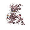
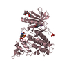
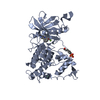
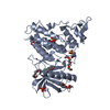
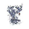
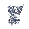
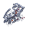
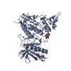
[English] 日本語
Yorodumi
- PDB-4yu2: Crystal structure of DYRK1A with harmine-derivatized AnnH-75 inhibitor -

+
Open data


ID or keywords:

Loading...

-
Basic information

Entry
Database: PDB / ID: 4yu2
TitleCrystal structure of DYRK1A with harmine-derivatized AnnH-75 inhibitor
ComponentsDual specificity tyrosine-phosphorylation-regulated kinase 1ADYRK1A
KeywordsTRANSFERASE / DYRK1A / down syndrome / inhibitor / harmine / Structural Genomics / Structural Genomics Consortium / SGC
Function / homology
Function and homology information


histone H3T45 kinase activity / positive regulation of protein deacetylation / peptidyl-serine autophosphorylation / negative regulation of DNA methylation-dependent heterochromatin formation / dual-specificity kinase / [RNA-polymerase]-subunit kinase / negative regulation of microtubule polymerization / tau-protein kinase activity / negative regulation of mRNA splicing, via spliceosome / negative regulation of DNA damage response, signal transduction by p53 class mediator ...histone H3T45 kinase activity / positive regulation of protein deacetylation / peptidyl-serine autophosphorylation / negative regulation of DNA methylation-dependent heterochromatin formation / dual-specificity kinase / [RNA-polymerase]-subunit kinase / negative regulation of microtubule polymerization / tau-protein kinase activity / negative regulation of mRNA splicing, via spliceosome / negative regulation of DNA damage response, signal transduction by p53 class mediator / amyloid-beta formation / G0 and Early G1 / peptidyl-tyrosine autophosphorylation / cytoskeletal protein binding / protein serine/threonine/tyrosine kinase activity / tubulin binding / RNA polymerase II CTD heptapeptide repeat kinase activity / positive regulation of RNA splicing / peptidyl-threonine phosphorylation / tau protein binding / non-membrane spanning protein tyrosine kinase activity / circadian rhythm / peptidyl-tyrosine phosphorylation / : / nervous system development / actin binding / peptidyl-serine phosphorylation / protein tyrosine kinase activity / protein autophosphorylation / transcription coactivator activity / cytoskeleton / protein kinase activity / nuclear speck / ribonucleoprotein complex / axon / protein phosphorylation / protein serine kinase activity / protein serine/threonine kinase activity / dendrite / positive regulation of DNA-templated transcription / nucleoplasm / ATP binding / identical protein binding / nucleus / cytoplasm
Similarity search - Function
Dual specificity tyrosine-phosphorylation-regulated kinase 1A/1B, catalytic domain / Transferase(Phosphotransferase) domain 1 / Transferase(Phosphotransferase); domain 1 / Phosphorylase Kinase; domain 1 / Phosphorylase Kinase; domain 1 / Serine/threonine-protein kinase, active site / Serine/Threonine protein kinases active-site signature. / Protein kinase domain / Serine/Threonine protein kinases, catalytic domain / Protein kinase, ATP binding site ...Dual specificity tyrosine-phosphorylation-regulated kinase 1A/1B, catalytic domain / Transferase(Phosphotransferase) domain 1 / Transferase(Phosphotransferase); domain 1 / Phosphorylase Kinase; domain 1 / Phosphorylase Kinase; domain 1 / Serine/threonine-protein kinase, active site / Serine/Threonine protein kinases active-site signature. / Protein kinase domain / Serine/Threonine protein kinases, catalytic domain / Protein kinase, ATP binding site / Protein kinases ATP-binding region signature. / Protein kinase domain profile. / Protein kinase domain / Protein kinase-like domain superfamily / 2-Layer Sandwich / Orthogonal Bundle / Mainly Alpha / Alpha Beta
Similarity search - Domain/homology
Chem-4H5 / Dual specificity tyrosine-phosphorylation-regulated kinase 1A
Similarity search - Component
Biological speciesHomo sapiens (human)
MethodX-RAY DIFFRACTION / SYNCHROTRON / MOLECULAR REPLACEMENT / Resolution: 2.9 Å
AuthorsChaikuad, A. / Wurzlbauer, A. / Nowak, R. / von Delft, F. / Arrowsmith, C.H. / Edwards, A.M. / Bountra, C. / Bracher, F. / Knapp, S. / Structural Genomics Consortium (SGC)
CitationJournal: Molecules / Year: 2020
Title: How to Separate Kinase Inhibition from Undesired Monoamine Oxidase A Inhibition-The Development of the DYRK1A Inhibitor AnnH75 from the Alkaloid Harmine.
Authors: Wurzlbauer, A. / Ruben, K. / Gurdal, E. / Chaikuad, A. / Knapp, S. / Sippl, W. / Becker, W. / Bracher, F.
History
DepositionMar 18, 2015Deposition site: RCSB / Processing site: PDBE
Revision 1.0Mar 25, 2015Provider: repository / Type: Initial release
Revision 1.1Jan 13, 2021Group: Database references / Category: citation / citation_author
Item: _citation.country / _citation.journal_abbrev ..._citation.country / _citation.journal_abbrev / _citation.journal_id_CSD / _citation.journal_id_ISSN / _citation.journal_volume / _citation.pdbx_database_id_DOI / _citation.pdbx_database_id_PubMed / _citation.title / _citation.year
Revision 1.2Jan 10, 2024Group: Data collection / Database references / Refinement description
Category: chem_comp_atom / chem_comp_bond ...chem_comp_atom / chem_comp_bond / database_2 / pdbx_initial_refinement_model / struct_ncs_dom_lim
Item: _database_2.pdbx_DOI / _database_2.pdbx_database_accession ..._database_2.pdbx_DOI / _database_2.pdbx_database_accession / _struct_ncs_dom_lim.beg_auth_comp_id / _struct_ncs_dom_lim.beg_label_asym_id / _struct_ncs_dom_lim.beg_label_comp_id / _struct_ncs_dom_lim.beg_label_seq_id / _struct_ncs_dom_lim.end_auth_comp_id / _struct_ncs_dom_lim.end_label_asym_id / _struct_ncs_dom_lim.end_label_comp_id / _struct_ncs_dom_lim.end_label_seq_id

-
Structure visualization

Structure viewerMolecule:
MolmilJmol/JSmol

Downloads & links

-
Assembly

Deposited unit
A: Dual specificity tyrosine-phosphorylation-regulated kinase 1A
B: Dual specificity tyrosine-phosphorylation-regulated kinase 1A
C: Dual specificity tyrosine-phosphorylation-regulated kinase 1A
D: Dual specificity tyrosine-phosphorylation-regulated kinase 1A
hetero molecules


Theoretical massNumber of molelcules
Total (without water)170,38015
Polymers168,3264
Non-polymers2,05411
Water5,513306
1
A: Dual specificity tyrosine-phosphorylation-regulated kinase 1A
hetero molecules


Theoretical massNumber of molelcules
Total (without water)42,5473
Polymers42,0821
Non-polymers4662
Water181
TypeNameSymmetry operationNumber
identity operation1_555x,y,z1
2
B: Dual specificity tyrosine-phosphorylation-regulated kinase 1A
hetero molecules


Theoretical massNumber of molelcules
Total (without water)42,8366
Polymers42,0821
Non-polymers7545
Water181
TypeNameSymmetry operationNumber
identity operation1_555x,y,z1
3
C: Dual specificity tyrosine-phosphorylation-regulated kinase 1A
hetero molecules


Theoretical massNumber of molelcules
Total (without water)42,6444
Polymers42,0821
Non-polymers5623
Water181
TypeNameSymmetry operationNumber
identity operation1_555x,y,z1
4
D: Dual specificity tyrosine-phosphorylation-regulated kinase 1A
hetero molecules


Theoretical massNumber of molelcules
Total (without water)42,3532
Polymers42,0821
Non-polymers2721
Water181
TypeNameSymmetry operationNumber
identity operation1_555x,y,z1
Unit cell
Length a, b, c (Å)265.567, 65.444, 138.648
Angle α, β, γ (deg.)90.00, 114.44, 90.00
Int Tables number5
Space group name H-MC121
Components on special symmetry positions
IDModelComponents
11A-612-

HOH

Noncrystallographic symmetry (NCS)NCS domain:
IDEns-IDDetails
11A
21B
12A
22C
13A
23D
14B
24C
15B
25D
16C
26D

NCS domain segments:

Component-ID: 0 / End auth comp-ID: LYS / End label comp-ID: LYS / Refine code: 0

Dom-IDEns-IDBeg auth comp-IDBeg label comp-IDAuth asym-IDLabel asym-IDAuth seq-IDLabel seq-ID
11LYSLYSAA134 - 48010 - 356
21LYSLYSBB134 - 48010 - 356
12VALVALAA135 - 48011 - 356
22VALVALCC135 - 48011 - 356
13VALVALAA135 - 48011 - 356
23VALVALDD135 - 48011 - 356
14VALVALBB135 - 48011 - 356
24VALVALCC135 - 48011 - 356
15VALVALBB135 - 48011 - 356
25VALVALDD135 - 48011 - 356
16VALVALCC135 - 48111 - 357
26VALVALDD135 - 48111 - 357

NCS ensembles :
ID
1
2
3
4
5
6

-
Components

#1: Protein
Dual specificity tyrosine-phosphorylation-regulated kinase 1A / DYRK1A / Dual specificity YAK1-related kinase / HP86 / Protein kinase minibrain homolog / hMNB


Mass: 42081.535 Da / Num. of mol.: 4 / Fragment: UNP residues 127-485
Source method: isolated from a genetically manipulated source
Details: kinase domain 127-485 / Source: (gene. exp.) Homo sapiens (human) / Gene: DYRK1A, DYRK, MNB, MNBH / Plasmid: pNIC28-Bsa4 / Details (production host): pNIC28-Bsa4 / Production host: Escherichia coli BL21(DE3) (bacteria) / Variant (production host): R3-pRARE2 / References: UniProt: Q13627, dual-specificity kinase
#2: Chemical ChemComp-PG4 / TETRAETHYLENE GLYCOL / Polyethylene glycol


Mass: 194.226 Da / Num. of mol.: 3 / Source method: obtained synthetically / Formula: C8H18O5 / Comment: precipitant*YM
#3: Chemical
ChemComp-4H5 / (1-chloro-7-methoxy-9H-beta-carbolin-9-yl)acetonitrile


Mass: 271.702 Da / Num. of mol.: 4 / Source method: obtained synthetically / Formula: C14H10ClN3O
#4: Chemical
ChemComp-SO4 / SULFATE ION / Sulfate


Mass: 96.063 Da / Num. of mol.: 4 / Source method: obtained synthetically / Formula: SO4
#5: Water ChemComp-HOH / water / Water


Mass: 18.015 Da / Num. of mol.: 306 / Source method: isolated from a natural source / Formula: H2O

-
Experimental details

-
Experiment

ExperimentMethod: X-RAY DIFFRACTION

-
Sample preparation

CrystalDensity Matthews: 3.26 Å3/Da / Density % sol: 62.25 %
Crystal growTemperature: 277 K / Method: vapor diffusion, sitting drop / pH: 9
Details: 31% PEG 400, 0.2 M lithium sulfate, 0.1 M Tris, pH 9.0

-
Data collection

DiffractionMean temperature: 100 K
Diffraction sourceSource: SYNCHROTRON / Site: Diamond / Beamline: I04-1 / Wavelength: 0.92 Å
DetectorType: DECTRIS PILATUS 2M / Detector: PIXEL / Date: Oct 31, 2013
RadiationMonochromator: Si (111) double crystal / Protocol: SINGLE WAVELENGTH / Monochromatic (M) / Laue (L): M / Scattering type: x-ray
Radiation wavelengthWavelength: 0.92 Å / Relative weight: 1
ReflectionResolution: 2.9→48.34 Å / Num. obs: 48349 / % possible obs: 99.5 % / Redundancy: 6.7 % / Biso Wilson estimate: 58.2 Å2 / Rmerge(I) obs: 0.126 / Net I/σ(I): 10.6
Reflection shellResolution: 2.9→3.06 Å / Redundancy: 6.8 % / Rmerge(I) obs: 0.928 / Mean I/σ(I) obs: 2 / % possible all: 99.5

-
Processing

Software
NameVersionClassification
REFMAC5.8.0049refinement
XDSdata reduction
SCALAdata scaling
PHASERphasing
RefinementMethod to determine structure: MOLECULAR REPLACEMENT
Starting model: 2WO6
Resolution: 2.9→48.34 Å / Cor.coef. Fo:Fc: 0.922 / Cor.coef. Fo:Fc free: 0.901 / SU B: 47.572 / SU ML: 0.44 / Cross valid method: THROUGHOUT / ESU R Free: 0.415 / Stereochemistry target values: MAXIMUM LIKELIHOOD / Details: HYDROGENS HAVE BEEN ADDED IN THE RIDING POSITIONS
RfactorNum. reflection% reflectionSelection details
Rfree0.27715 2443 5.1 %RANDOM
Rwork0.24302 ---
obs0.24478 45905 99.34 %-
Solvent computationIon probe radii: 0.8 Å / Shrinkage radii: 0.8 Å / VDW probe radii: 1.2 Å / Solvent model: MASK
Displacement parametersBiso mean: 75.108 Å2
Baniso -1Baniso -2Baniso -3
1-0.28 Å2-0 Å2-2.92 Å2
2---2.02 Å20 Å2
3---3.12 Å2
Refine analyzeLuzzati coordinate error obs: 0.515 Å
Refinement stepCycle: 1 / Resolution: 2.9→48.34 Å
ProteinNucleic acidLigandSolventTotal
Num. atoms11253 0 135 306 11694
Refine LS restraints
Refine-IDTypeDev idealDev ideal targetNumber
X-RAY DIFFRACTIONr_bond_refined_d0.0110.01911735
X-RAY DIFFRACTIONr_bond_other_d0.0050.0211117
X-RAY DIFFRACTIONr_angle_refined_deg1.2231.98615871
X-RAY DIFFRACTIONr_angle_other_deg0.825325541
X-RAY DIFFRACTIONr_dihedral_angle_1_deg6.70851399
X-RAY DIFFRACTIONr_dihedral_angle_2_deg36.76823.768552
X-RAY DIFFRACTIONr_dihedral_angle_3_deg15.426152039
X-RAY DIFFRACTIONr_dihedral_angle_4_deg19.9911576
X-RAY DIFFRACTIONr_chiral_restr0.0630.21672
X-RAY DIFFRACTIONr_gen_planes_refined0.0070.02113121
X-RAY DIFFRACTIONr_gen_planes_other0.0050.022795
X-RAY DIFFRACTIONr_nbd_refined
X-RAY DIFFRACTIONr_nbd_other
X-RAY DIFFRACTIONr_nbtor_refined
X-RAY DIFFRACTIONr_nbtor_other
X-RAY DIFFRACTIONr_xyhbond_nbd_refined
X-RAY DIFFRACTIONr_xyhbond_nbd_other
X-RAY DIFFRACTIONr_metal_ion_refined
X-RAY DIFFRACTIONr_metal_ion_other
X-RAY DIFFRACTIONr_symmetry_vdw_refined
X-RAY DIFFRACTIONr_symmetry_vdw_other
X-RAY DIFFRACTIONr_symmetry_hbond_refined
X-RAY DIFFRACTIONr_symmetry_hbond_other
X-RAY DIFFRACTIONr_symmetry_metal_ion_refined
X-RAY DIFFRACTIONr_symmetry_metal_ion_other
X-RAY DIFFRACTIONr_mcbond_it1.9843.1815576
X-RAY DIFFRACTIONr_mcbond_other1.9843.1825577
X-RAY DIFFRACTIONr_mcangle_it3.4014.7666961
X-RAY DIFFRACTIONr_mcangle_other3.44.7676962
X-RAY DIFFRACTIONr_scbond_it2.4533.3766159
X-RAY DIFFRACTIONr_scbond_other2.3993.3586143
X-RAY DIFFRACTIONr_scangle_it
X-RAY DIFFRACTIONr_scangle_other3.6584.998881
X-RAY DIFFRACTIONr_long_range_B_refined7.57527.31413723
X-RAY DIFFRACTIONr_long_range_B_other7.57427.31913724
X-RAY DIFFRACTIONr_rigid_bond_restr
X-RAY DIFFRACTIONr_sphericity_free
X-RAY DIFFRACTIONr_sphericity_bonded
Refine LS restraints NCS

Refine-ID: X-RAY DIFFRACTION / Type: interatomic distance / Weight position: 0.05

Ens-IDDom-IDAuth asym-IDNumberRms dev position (Å)
11A221010.05
12B221010.05
21A221870.05
22C221870.05
31A221150.05
32D221150.05
41B220410.04
42C220410.04
51B220440.04
52D220440.04
61C219880.05
62D219880.05
LS refinement shellResolution: 2.9→2.975 Å / Total num. of bins used: 20
RfactorNum. reflection% reflection
Rfree0.419 197 -
Rwork0.393 3358 -
obs--99.38 %
Refinement TLS params.

Method: refined / Refine-ID: X-RAY DIFFRACTION

IDL112)L122)L132)L222)L232)L332)S11 (Å °)S12 (Å °)S13 (Å °)S21 (Å °)S22 (Å °)S23 (Å °)S31 (Å °)S32 (Å °)S33 (Å °)T112)T122)T132)T222)T232)T332)Origin x (Å)Origin y (Å)Origin z (Å)
14.01320.57421.76671.49910.21611.50920.2455-0.2724-0.2672-0.0262-0.15310.15150.1983-0.3516-0.09240.2929-0.12140.14880.59540.03840.1735-41.2569-43.64722.0025
25.6909-1.10050.86015.40570.74063.50940.1308-0.1591-0.48070.14480.03-0.6068-0.00590.2581-0.16070.4511-0.13770.10280.4722-0.08640.187711.8262-16.469131.8933
32.75110.69481.32841.65350.85393.4960.1997-0.1965-0.01920.3123-0.18930.06360.0004-0.2649-0.01050.2055-0.04290.08930.22030.00090.0491-5.9335-27.770715.4181
41.7315-0.3068-1.00661.79160.71373.4025-0.0444-0.2230.117-0.27950.06920.1021-0.4671-0.2056-0.02480.5846-0.03890.10810.5946-0.08630.0578-4.8271-56.922656.5865
51.40990.03550.11561.72470.69563.8811-0.08910.14750.19110.0969-0.01030.1987-0.75530.04980.09940.4664-0.08310.04780.62110.21940.2339-66.381-51.422133.1828
Refinement TLS group
IDRefine-IDRefine TLS-IDAuth asym-IDAuth seq-ID
1X-RAY DIFFRACTION1B134 - 481
2X-RAY DIFFRACTION2A134 - 219
3X-RAY DIFFRACTION3A220 - 482
4X-RAY DIFFRACTION4C135 - 481
5X-RAY DIFFRACTION5D135 - 481

+
About Yorodumi

-
News

-
Feb 9, 2022. New format data for meta-information of EMDB entries

New format data for meta-information of EMDB entries

  • Version 3 of the EMDB header file is now the official format.
  • The previous official version 1.9 will be removed from the archive.

Related info.:EMDB header

External links:wwPDB to switch to version 3 of the EMDB data model

-
Aug 12, 2020. Covid-19 info

Covid-19 info

URL: https://pdbj.org/emnavi/covid19.php

New page: Covid-19 featured information page in EM Navigator.

Related info.:Covid-19 info / Mar 5, 2020. Novel coronavirus structure data

+
Mar 5, 2020. Novel coronavirus structure data

Novel coronavirus structure data

Related info.:Yorodumi Speices / Aug 12, 2020. Covid-19 info

External links:COVID-19 featured content - PDBj / Molecule of the Month (242):Coronavirus Proteases

+
Jan 31, 2019. EMDB accession codes are about to change! (news from PDBe EMDB page)

EMDB accession codes are about to change! (news from PDBe EMDB page)

  • The allocation of 4 digits for EMDB accession codes will soon come to an end. Whilst these codes will remain in use, new EMDB accession codes will include an additional digit and will expand incrementally as the available range of codes is exhausted. The current 4-digit format prefixed with “EMD-” (i.e. EMD-XXXX) will advance to a 5-digit format (i.e. EMD-XXXXX), and so on. It is currently estimated that the 4-digit codes will be depleted around Spring 2019, at which point the 5-digit format will come into force.
  • The EM Navigator/Yorodumi systems omit the EMD- prefix.

Related info.:Q: What is EMD? / ID/Accession-code notation in Yorodumi/EM Navigator

External links:EMDB Accession Codes are Changing Soon! / Contact to PDBj

+
Jul 12, 2017. Major update of PDB

Major update of PDB

  • wwPDB released updated PDB data conforming to the new PDBx/mmCIF dictionary.
  • This is a major update changing the version number from 4 to 5, and with Remediation, in which all the entries are updated.
  • In this update, many items about electron microscopy experimental information are reorganized (e.g. em_software).
  • Now, EM Navigator and Yorodumi are based on the updated data.

External links:wwPDB Remediation / Enriched Model Files Conforming to OneDep Data Standards Now Available in the PDB FTP Archive

-
Yorodumi

Thousand views of thousand structures

  • Yorodumi is a browser for structure data from EMDB, PDB, SASBDB, etc.
  • This page is also the successor to EM Navigator detail page, and also detail information page/front-end page for Omokage search.
  • The word "yorodu" (or yorozu) is an old Japanese word meaning "ten thousand". "mi" (miru) is to see.

Related info.:EMDB / PDB / SASBDB / Comparison of 3 databanks / Yorodumi Search / Aug 31, 2016. New EM Navigator & Yorodumi / Yorodumi Papers / Jmol/JSmol / Function and homology information / Changes in new EM Navigator and Yorodumi

Read more