 Movie
Movie Controller


+ Open data

- Basic information
| Entry | Database: PDB / ID: 1ikq | ||||||
|---|---|---|---|---|---|---|---|
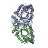
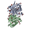
| Title | Pseudomonas Aeruginosa Exotoxin A, wild type | ||||||
 Components Components | EXOTOXIN A | ||||||
 Keywords Keywords | TRANSFERASE / DOMAIN I / II / III of EXOTOXIN A | ||||||
| Function / homology |  Function and homology information Function and homology informationsymbiont-mediated suppression of host translation elongation / NAD+-diphthamide ADP-ribosyltransferase / NAD+-diphthamide ADP-ribosyltransferase activity / symbiont-mediated killing of host cell / nucleotidyltransferase activity / toxin activity Similarity search - Function | ||||||
| Biological species |   Pseudomonas aeruginosa (bacteria) Pseudomonas aeruginosa (bacteria) | ||||||
| Method |  X-RAY DIFFRACTION / SYNCHROTRON / MOLECULAR REPLACEMENT / Resolution: 1.62 Å X-RAY DIFFRACTION / SYNCHROTRON / MOLECULAR REPLACEMENT / Resolution: 1.62 Å | ||||||
 Authors Authors | McKay, D.B. / Wedekind, J.E. / Trame, C.B. | ||||||
 Citation Citation | Journal: J.Mol.Biol. / Year: 2001 Title: Refined Crystallographic Structure of Pseudomonas aeruginosa Exotoxin A and its Implications for the Molecular Mechanism of Toxicity Authors: Wedekind, J.E. / Trame, C.B. / Dorywalska, M. / Koehl, P. / Raschke, T.M. / McKee, M. / FitzGerald, D. / Collier, R.J. / McKay, D.B. #1: Journal: J.Mol.Biol. / Year: 1982Title: Crystallization of Exotoxin A from Pseudomonas aeruginosa Authors: Collier, R.J. / McKay, D.B. #2: Journal: Proc.Natl.Acad.Sci.USA / Year: 1986Title: Structure of Exotoxin A of Pseudomonas aeruginosa at 3.0-Angstrom Resolution Authors: Allured, V.S. / Collier, R.J. / Carroll, S.F. / McKay, D.B. #3: Journal: Proteins / Year: 1988Title: Mapping the Enzymatic Active Site of Pseudomonas aeruginosa Exotoxin A Authors: Brandhuber, B.J. / Allured, V.S. / G., Falbel T. / B., McKay D. #4: Journal: Proc.Natl.Acad.Sci.USA / Year: 1995Title: The Crystal Structure of Pseudomonas aeruginosa Exotoxin Domain III with Nicotinamide and AMP: conformational differences with the intact exotoxin Authors: Li, M. / Dyda, F. / Benhar, I. / Pastan, I. / Davies, D.R. | ||||||
| History |
|
- Structure visualization
| Structure viewer | Molecule: MolmilJmol/JSmol |
|---|
- Downloads & links
-Download
| PDBx/mmCIF format | 1ikq.cif.gz | 141.5 KB | Display | PDBx/mmCIF format |
|---|---|---|---|---|
| PDB format | pdb1ikq.ent.gz | 108.7 KB | Display | PDB format |
| PDBx/mmJSON format | 1ikq.json.gz | Tree view | PDBx/mmJSON format | |
| Others |  Other downloads Other downloads |
-Validation report
| Arichive directory | https://data.pdbj.org/pub/pdb/validation_reports/ik/1ikqftp://data.pdbj.org/pub/pdb/validation_reports/ik/1ikq | HTTPS FTP |
|---|
-Related structure data


-Links
 PDBj
PDBj
- Assembly
| Deposited unit | 
| ||||||||
|---|---|---|---|---|---|---|---|---|---|
| 1 |
| ||||||||
| Unit cell |
|
-Components
| #1: Protein | Mass: 66871.938 Da / Num. of mol.: 1 Source method: isolated from a genetically manipulated source Source: (gene. exp.) Pseudomonas aeruginosa (bacteria) / Gene: PE / Plasmid: pMOA1A2VK352 f+ T / Species (production host): Escherichia coli / Production host: References: UniProt: P11439, Transferases; Glycosyltransferases; Pentosyltransferases | ||||||
|---|---|---|---|---|---|---|---|
| #2: Chemical |   Mass: 35.453 Da / Num. of mol.: 2 / Source method: obtained synthetically / Formula: Cl Mass: 35.453 Da / Num. of mol.: 2 / Source method: obtained synthetically / Formula: Cl#3: Chemical |   Mass: 22.990 Da / Num. of mol.: 2 / Source method: obtained synthetically / Formula: Na Mass: 22.990 Da / Num. of mol.: 2 / Source method: obtained synthetically / Formula: Na#4: Water | ChemComp-HOH / |  Mass: 18.015 Da / Num. of mol.: 546 / Source method: isolated from a natural source / Formula: H2O Mass: 18.015 Da / Num. of mol.: 546 / Source method: isolated from a natural source / Formula: H2OHas protein modification | Y | |
-Experimental details
-Experiment
| Experiment | Method: X-RAY DIFFRACTION / Number of used crystals: 2 |
|---|
- Sample preparation
| Crystal | Density Matthews: 2.52 Å3/Da / Density % sol: 51.23 % |
|---|---|
| Crystal grow | Temperature: 281 K / Method: vapor diffusion, hanging drop / pH: 8 Details: PEG 8000, sodium chloride, hepes, pH 8.0, VAPOR DIFFUSION, HANGING DROP, temperature 281K |
| Crystal grow | *PLUS Details: Collier, R.J., (1982) J. Mol. Biol., 157, 413. |
-Data collection
| Diffraction |
| ||||||||||||||||||
|---|---|---|---|---|---|---|---|---|---|---|---|---|---|---|---|---|---|---|---|
| Diffraction source |
| ||||||||||||||||||
| Detector |
| ||||||||||||||||||
| Radiation |
| ||||||||||||||||||
| Radiation wavelength |
| ||||||||||||||||||
| Reflection | Resolution: 1.62→50 Å / Observed criterion σ(F): 2 / Observed criterion σ(I): 2 / Biso Wilson estimate: 21.4 Å2 | ||||||||||||||||||
| Reflection | *PLUS Lowest resolution: 20 Å / Num. obs: 80328 / % possible obs: 93 % / Num. measured all: 224879 / Rmerge(I) obs: 0.04 | ||||||||||||||||||
| Reflection shell | *PLUS Highest resolution: 1.62 Å / Lowest resolution: 1.71 Å / % possible obs: 91 % / Rmerge(I) obs: 0.185 |

- Processing
| Software |
| ||||||||||||||||||||||||||||||||||||||||||||
|---|---|---|---|---|---|---|---|---|---|---|---|---|---|---|---|---|---|---|---|---|---|---|---|---|---|---|---|---|---|---|---|---|---|---|---|---|---|---|---|---|---|---|---|---|---|
| Refinement | Method to determine structure: MOLECULAR REPLACEMENT Starting model: 3A EXOTOXIN A model (ref. 3) Resolution: 1.62→49.79 Å / Rfactor Rfree error: 0.003 / Data cutoff high absF: 1832154.15 / Data cutoff low absF: 0 / Isotropic thermal model: RESTRAINED / Cross valid method: THROUGHOUT / σ(F): 0 / σ(I): 0 / Stereochemistry target values: Engh & Huber
| ||||||||||||||||||||||||||||||||||||||||||||
| Solvent computation | Solvent model: FLAT MODEL / Bsol: 54.8 Å2 / ksol: 0.369 e/Å3 | ||||||||||||||||||||||||||||||||||||||||||||
| Displacement parameters | Biso mean: 27.5 Å2
| ||||||||||||||||||||||||||||||||||||||||||||
| Refine analyze |
| ||||||||||||||||||||||||||||||||||||||||||||
| Refinement step | Cycle: LAST / Resolution: 1.62→49.79 Å
| ||||||||||||||||||||||||||||||||||||||||||||
| Refine LS restraints |
| ||||||||||||||||||||||||||||||||||||||||||||
| LS refinement shell | Resolution: 1.62→1.69 Å / Rfactor Rfree error: 0.012 / Total num. of bins used: 8
| ||||||||||||||||||||||||||||||||||||||||||||
| Xplor file |
| ||||||||||||||||||||||||||||||||||||||||||||
| Software | *PLUS Name: CNS / Version: 1 / Classification: refinement | ||||||||||||||||||||||||||||||||||||||||||||
| Refinement | *PLUS Lowest resolution: 50 Å / Num. reflection obs: 73810 / σ(F): 0 / % reflection Rfree: 8.1 % | ||||||||||||||||||||||||||||||||||||||||||||
| Solvent computation | *PLUS | ||||||||||||||||||||||||||||||||||||||||||||
| Displacement parameters | *PLUS Biso mean: 27.5 Å2 | ||||||||||||||||||||||||||||||||||||||||||||
| Refine LS restraints | *PLUS
| ||||||||||||||||||||||||||||||||||||||||||||
| LS refinement shell | *PLUS Rfactor Rfree: 0.32 / % reflection Rfree: 7.8 % / Rfactor Rwork: 0.286 / Rfactor obs: 0.286 |
