 Movie
Movie Controller

[English] 日本語
 Yorodumi
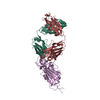
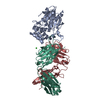
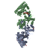
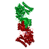
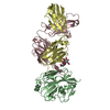
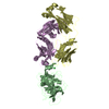
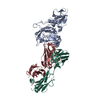
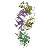
- PDB-1dma: DOMAIN III OF PSEUDOMONAS AERUGINOSA EXOTOXIN COMPLEXED WITH NICO... -
+ Open data

- Basic information
| Entry | Database: PDB / ID: 1dma | ||||||
|---|---|---|---|---|---|---|---|
| Title | DOMAIN III OF PSEUDOMONAS AERUGINOSA EXOTOXIN COMPLEXED WITH NICOTINAMIDE AND AMP | ||||||
 Components Components | EXOTOXIN A | ||||||
 Keywords Keywords | ADP-RIBOSYLATION | ||||||
| Function / homology |  Function and homology information Function and homology informationsymbiont-mediated suppression of host translation elongation / NAD+-diphthamide ADP-ribosyltransferase / NAD+-diphthamide ADP-ribosyltransferase activity / symbiont-mediated killing of host cell / nucleotidyltransferase activity / toxin activity Similarity search - Function | ||||||
| Biological species |   Pseudomonas aeruginosa (bacteria) Pseudomonas aeruginosa (bacteria) | ||||||
| Method |  X-RAY DIFFRACTION / Resolution: 2.5 Å X-RAY DIFFRACTION / Resolution: 2.5 Å | ||||||
 Authors Authors | Li, M. / Dyda, F. / Benhar, I. / Pastan, I. / Davies, D. | ||||||
 Citation Citation | Journal: Proc.Natl.Acad.Sci.USA / Year: 1995 Title: The crystal structure of Pseudomonas aeruginosa exotoxin domain III with nicotinamide and AMP: conformational differences with the intact exotoxin. Authors: Li, M. / Dyda, F. / Benhar, I. / Pastan, I. / Davies, D.R. #1: Journal: J.Biol.Chem. / Year: 1989Title: Functional Analysis of Domains II, Ib, and III of Pseudomonas Exotoxin Authors: Siegall, C.B. / Chaudhary, V.K. / Fitzgerald, D.J. / Pastan, I. #2: Journal: Proc.Natl.Acad.Sci.USA / Year: 1986Title: Structure of Exotoxin a of Pseudomonas Aeruginosa a 3.0-Angstrom Resolution Authors: Allured, V.S. / Collier, R.J. / Carroll, S.F. / Mckay, D.B. | ||||||
| History |
|
- Structure visualization
| Structure viewer | Molecule: MolmilJmol/JSmol |
|---|
- Downloads & links
-Download
| PDBx/mmCIF format | 1dma.cif.gz | 88.9 KB | Display | PDBx/mmCIF format |
|---|---|---|---|---|
| PDB format | pdb1dma.ent.gz | 68 KB | Display | PDB format |
| PDBx/mmJSON format | 1dma.json.gz | Tree view | PDBx/mmJSON format | |
| Others |  Other downloads Other downloads |
-Validation report
| Arichive directory | https://data.pdbj.org/pub/pdb/validation_reports/dm/1dmaftp://data.pdbj.org/pub/pdb/validation_reports/dm/1dma | HTTPS FTP |
|---|
-Related structure data
| Similar structure data |
|---|

-Links
 PDBj
PDBj


- Assembly
| Deposited unit | 
| ||||||||
|---|---|---|---|---|---|---|---|---|---|
| 1 | 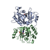
| ||||||||
| Unit cell |
| ||||||||
| Atom site foot note | 1: CIS PROLINE - PRO A 536 / 2: CIS PROLINE - PRO B 536 |
-Components
| #1: Protein | Mass: 23334.041 Da / Num. of mol.: 2 Source method: isolated from a genetically manipulated source Source: (gene. exp.) Pseudomonas aeruginosa (bacteria) / Plasmid: PPED5-399 / Production host: #2: Chemical | 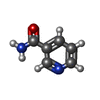  Mass: 122.125 Da / Num. of mol.: 2 / Source method: obtained synthetically / Formula: C6H6N2O / Comment: medication*YM Mass: 122.125 Da / Num. of mol.: 2 / Source method: obtained synthetically / Formula: C6H6N2O / Comment: medication*YM#3: Chemical | ChemComp-AMP / |   Mass: 347.221 Da / Num. of mol.: 1 / Source method: obtained synthetically / Formula: C10H14N5O7P / Comment: AMP*YM Mass: 347.221 Da / Num. of mol.: 1 / Source method: obtained synthetically / Formula: C10H14N5O7P / Comment: AMP*YM#4: Water | ChemComp-HOH / |  Mass: 18.015 Da / Num. of mol.: 33 / Source method: isolated from a natural source / Formula: H2O Mass: 18.015 Da / Num. of mol.: 33 / Source method: isolated from a natural source / Formula: H2O |
|---|
-Experimental details
-Experiment
| Experiment | Method: X-RAY DIFFRACTION |
|---|
- Sample preparation
| Crystal | Density Matthews: 2.75 Å3/Da / Density % sol: 55.24 % | ||||||||||||||||||||||||||||||||||||||||||||||||
|---|---|---|---|---|---|---|---|---|---|---|---|---|---|---|---|---|---|---|---|---|---|---|---|---|---|---|---|---|---|---|---|---|---|---|---|---|---|---|---|---|---|---|---|---|---|---|---|---|---|
| Crystal grow | *PLUS pH: 7 / Method: vapor diffusion, hanging drop / Details: used as seeds | ||||||||||||||||||||||||||||||||||||||||||||||||
| Components of the solutions | *PLUS
|
-Data collection
| Radiation | Scattering type: x-ray |
|---|---|
| Radiation wavelength | Relative weight: 1 |
| Reflection | Resolution: 2.5→8 Å / Num. obs: 16513 / % possible obs: 88.9 % / Observed criterion σ(I): 2 |
| Reflection | *PLUS Rmerge(I) obs: 0.068 |
- Processing
| Software | Name: X-PLOR / Version: 3.1 / Classification: refinement | ||||||||||||||||||||||||||||||||||||||||||||||||||||||||||||
|---|---|---|---|---|---|---|---|---|---|---|---|---|---|---|---|---|---|---|---|---|---|---|---|---|---|---|---|---|---|---|---|---|---|---|---|---|---|---|---|---|---|---|---|---|---|---|---|---|---|---|---|---|---|---|---|---|---|---|---|---|---|
| Refinement | Resolution: 2.5→8 Å / σ(F): 2
| ||||||||||||||||||||||||||||||||||||||||||||||||||||||||||||
| Displacement parameters | Biso mean: 32.3 Å2 | ||||||||||||||||||||||||||||||||||||||||||||||||||||||||||||
| Refine analyze | Luzzati coordinate error obs: 0.3 Å | ||||||||||||||||||||||||||||||||||||||||||||||||||||||||||||
| Refinement step | Cycle: LAST / Resolution: 2.5→8 Å
| ||||||||||||||||||||||||||||||||||||||||||||||||||||||||||||
| Refine LS restraints |
| ||||||||||||||||||||||||||||||||||||||||||||||||||||||||||||
| Software | *PLUS Version: 3.1 / Classification: refinement | ||||||||||||||||||||||||||||||||||||||||||||||||||||||||||||
| Refinement | *PLUS Rfactor Rfree: 0.264 | ||||||||||||||||||||||||||||||||||||||||||||||||||||||||||||
| Solvent computation | *PLUS | ||||||||||||||||||||||||||||||||||||||||||||||||||||||||||||
| Displacement parameters | *PLUS | ||||||||||||||||||||||||||||||||||||||||||||||||||||||||||||
| Refine LS restraints | *PLUS
|
