 Movie
Movie Controller

[English] 日本語
 Yorodumi
- PDB-1aer: DOMAIN III OF PSEUDOMONAS AERUGINOSA EXOTOXIN COMPLEXED WITH BETA-TAD -
+ Open data

- Basic information
| Entry | Database: PDB / ID: 1aer | ||||||
|---|---|---|---|---|---|---|---|
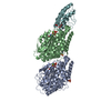
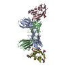
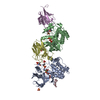
| Title | DOMAIN III OF PSEUDOMONAS AERUGINOSA EXOTOXIN COMPLEXED WITH BETA-TAD | ||||||
 Components Components | EXOTOXIN A | ||||||
 Keywords Keywords | ADP-RIBOSYLATION / TOXIN / TRANSFERASE / GLYCOSYLTRANSFERASE / NAD | ||||||
| Function / homology |  Function and homology information Function and homology informationsymbiont-mediated suppression of host translation elongation / NAD+-diphthamide ADP-ribosyltransferase / NAD+-diphthamide ADP-ribosyltransferase activity / symbiont-mediated killing of host cell / nucleotidyltransferase activity / toxin activity Similarity search - Function | ||||||
| Biological species |   Pseudomonas aeruginosa (bacteria) Pseudomonas aeruginosa (bacteria) | ||||||
| Method |  X-RAY DIFFRACTION / Resolution: 2.3 Å X-RAY DIFFRACTION / Resolution: 2.3 Å | ||||||
 Authors Authors | Li, M. / Dyda, F. / Benhar, I. / Pastan, I. / Davies, D.R. | ||||||
 Citation Citation | Journal: Proc.Natl.Acad.Sci.USA / Year: 1996 Title: Crystal structure of the catalytic domain of Pseudomonas exotoxin A complexed with a nicotinamide adenine dinucleotide analog: implications for the activation process and for ADP ribosylation Authors: Li, M. / Dyda, F. / Benhar, I. / Pastan, I. / Davies, D.R. #1: Journal: Proc.Natl.Acad.Sci.USA / Year: 1995Title: The Crystal Structure of Pseudomonas Aeruginosa Exotoxin Domain III with Nicotinamide and AMP: Conformational Differences with the Intact Exotoxin Authors: Li, M. / Dyda, F. / Benhar, I. / Pastan, I. / Davies, D.R. #2: Journal: Proc.Natl.Acad.Sci.USA / Year: 1986Title: Structure of Exotoxin a of Pseudomonas Aeruginosa at 3.0-Angstrom Resolution Authors: Allured, V.S. / Collier, R.J. / Carroll, S.F. / Mckay, D.B. | ||||||
| History |
|
- Structure visualization
| Structure viewer | Molecule: MolmilJmol/JSmol |
|---|
- Downloads & links
-Download
| PDBx/mmCIF format | 1aer.cif.gz | 93.3 KB | Display | PDBx/mmCIF format |
|---|---|---|---|---|
| PDB format | pdb1aer.ent.gz | 70.5 KB | Display | PDB format |
| PDBx/mmJSON format | 1aer.json.gz | Tree view | PDBx/mmJSON format | |
| Others |  Other downloads Other downloads |
-Validation report
| Arichive directory | https://data.pdbj.org/pub/pdb/validation_reports/ae/1aerftp://data.pdbj.org/pub/pdb/validation_reports/ae/1aer | HTTPS FTP |
|---|
-Related structure data
| Similar structure data |
|---|

-Links
 PDBj
PDBj


- Assembly
| Deposited unit | 
| ||||||||
|---|---|---|---|---|---|---|---|---|---|
| 1 | 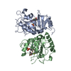
| ||||||||
| Unit cell |
|
-Components
| #1: Protein | Mass: 22918.580 Da / Num. of mol.: 2 / Fragment: DOMAIN III OF PSEUDOMONAS TOXIN Source method: isolated from a genetically manipulated source Source: (gene. exp.) Pseudomonas aeruginosa (bacteria) / Plasmid: PPED5-399 / Production host: References: UniProt: P11439, Transferases; Glycosyltransferases; Pentosyltransferases #2: Chemical | ChemComp-TAD / |   Mass: 667.480 Da / Num. of mol.: 1 / Source method: obtained synthetically / Formula: C20H27N7O13P2S Mass: 667.480 Da / Num. of mol.: 1 / Source method: obtained synthetically / Formula: C20H27N7O13P2S#3: Chemical | ChemComp-TIA / |   Mass: 244.268 Da / Num. of mol.: 1 Mass: 244.268 Da / Num. of mol.: 1Source method: isolated from a genetically manipulated source Formula: C9H12N2O4S #4: Chemical | ChemComp-AMP / |   Mass: 347.221 Da / Num. of mol.: 1 / Source method: obtained synthetically / Formula: C10H14N5O7P / Comment: AMP*YM Mass: 347.221 Da / Num. of mol.: 1 / Source method: obtained synthetically / Formula: C10H14N5O7P / Comment: AMP*YM#5: Water | ChemComp-HOH / |  Mass: 18.015 Da / Num. of mol.: 108 / Source method: isolated from a natural source / Formula: H2O Mass: 18.015 Da / Num. of mol.: 108 / Source method: isolated from a natural source / Formula: H2OHas protein modification | N | Sequence details | THE SEQUENCE IN THIS ENTRY IS FROM 400 TO 613 FROM THAT OF THE COMPLETE TOXIN IN GENE BANK. THERE ...THE SEQUENCE IN THIS ENTRY IS FROM 400 TO 613 FROM THAT OF THE COMPLETE TOXIN IN GENE BANK. THERE ARE TWO MONOMERS IN THE ASYMMETRIC | |
|---|
-Experimental details
-Experiment
| Experiment | Method: X-RAY DIFFRACTION |
|---|
- Sample preparation
| Crystal | Density Matthews: 2.8 Å3/Da / Density % sol: 50.2 % | |||||||||||||||||||||||||||||||||||||||||||||||||
|---|---|---|---|---|---|---|---|---|---|---|---|---|---|---|---|---|---|---|---|---|---|---|---|---|---|---|---|---|---|---|---|---|---|---|---|---|---|---|---|---|---|---|---|---|---|---|---|---|---|---|
| Crystal grow | *PLUS pH: 7 / Method: vapor diffusion, hanging drop | |||||||||||||||||||||||||||||||||||||||||||||||||
| Components of the solutions | *PLUS
|
-Data collection
| Diffraction | Mean temperature: 293 K |
|---|---|
| Diffraction source | Wavelength: 1.5418 |
| Detector | Type: RIGAKU RAXIS II / Detector: IMAGE PLATE / Date: Apr 1, 1995 |
| Radiation | Monochromatic (M) / Laue (L): M / Scattering type: x-ray |
| Radiation wavelength | Wavelength: 1.5418 Å / Relative weight: 1 |
| Reflection | Resolution: 2.3→50 Å / Num. obs: 22417 / Observed criterion σ(I): 2 / Redundancy: 5.1 % / Rmerge(I) obs: 0.057 |
| Reflection | *PLUS % possible obs: 93.4 % / Redundancy: 4.7 % |
| Reflection shell | *PLUS Highest resolution: 2.3 Å / Lowest resolution: 2.4 Å / % possible obs: 78.4 % |
- Processing
| Software |
| ||||||||||||||||||||||||||||||||||||||||||||||||||||||||||||
|---|---|---|---|---|---|---|---|---|---|---|---|---|---|---|---|---|---|---|---|---|---|---|---|---|---|---|---|---|---|---|---|---|---|---|---|---|---|---|---|---|---|---|---|---|---|---|---|---|---|---|---|---|---|---|---|---|---|---|---|---|---|
| Refinement | Resolution: 2.3→8 Å / σ(F): 2
| ||||||||||||||||||||||||||||||||||||||||||||||||||||||||||||
| Displacement parameters | Biso mean: 28.17 Å2 | ||||||||||||||||||||||||||||||||||||||||||||||||||||||||||||
| Refinement step | Cycle: LAST / Resolution: 2.3→8 Å
| ||||||||||||||||||||||||||||||||||||||||||||||||||||||||||||
| Refine LS restraints |
| ||||||||||||||||||||||||||||||||||||||||||||||||||||||||||||
| Software | *PLUS Name: X-PLOR / Classification: refinement | ||||||||||||||||||||||||||||||||||||||||||||||||||||||||||||
| Refinement | *PLUS | ||||||||||||||||||||||||||||||||||||||||||||||||||||||||||||
| Solvent computation | *PLUS | ||||||||||||||||||||||||||||||||||||||||||||||||||||||||||||
| Displacement parameters | *PLUS | ||||||||||||||||||||||||||||||||||||||||||||||||||||||||||||
| Refine LS restraints | *PLUS
|
