 Movie
Movie Controller


+ Open data

- Basic information
| Entry | Database: PDB / ID: 1gsf | ||||||
|---|---|---|---|---|---|---|---|
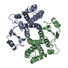
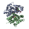
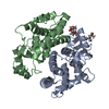
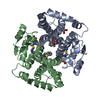
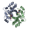
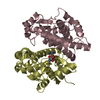
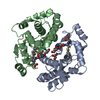
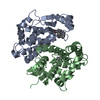
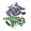
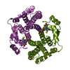
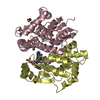
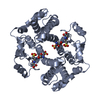
| Title | GLUTATHIONE TRANSFERASE A1-1 COMPLEXED WITH ETHACRYNIC ACID | ||||||
 Components Components | GLUTATHIONE TRANSFERASE A1-1 | ||||||
 Keywords Keywords | TRANSFERASE (GLUTATHIONE) / A1-1 | ||||||
| Function / homology |  Function and homology information Function and homology informationIsomerases; Intramolecular oxidoreductases; Transposing C=C bonds / glutathione derivative biosynthetic process / linoleic acid metabolic process / steroid Delta-isomerase activity / Glutathione conjugation / glutathione peroxidase activity / Azathioprine ADME / Heme degradation / NFE2L2 regulating anti-oxidant/detoxification enzymes / prostaglandin metabolic process ...Isomerases; Intramolecular oxidoreductases; Transposing C=C bonds / glutathione derivative biosynthetic process / linoleic acid metabolic process / steroid Delta-isomerase activity / Glutathione conjugation / glutathione peroxidase activity / Azathioprine ADME / Heme degradation / NFE2L2 regulating anti-oxidant/detoxification enzymes / prostaglandin metabolic process / glutathione transferase / glutathione transferase activity / Oxidoreductases; Acting on a peroxide as acceptor; Peroxidases / epithelial cell differentiation / glutathione metabolic process / xenobiotic metabolic process / fatty acid binding / extracellular exosome / cytosol Similarity search - Function | ||||||
| Biological species |  Homo sapiens (human) Homo sapiens (human) | ||||||
| Method |  X-RAY DIFFRACTION / Resolution: 2.7 Å X-RAY DIFFRACTION / Resolution: 2.7 Å | ||||||
 Authors Authors | L'Hermite, G. / Sinning, I. / Cameron, A.D. / Jones, T.A. | ||||||
 Citation Citation | Journal: Structure / Year: 1995 Title: Structural analysis of human alpha-class glutathione transferase A1-1 in the apo-form and in complexes with ethacrynic acid and its glutathione conjugate. Authors: Cameron, A.D. / Sinning, I. / L'Hermite, G. / Olin, B. / Board, P.G. / Mannervik, B. / Jones, T.A. #1: Journal: J.Mol.Biol. / Year: 1993Title: Structure Determination and Refinement of Human Alpha Class Glutathione Transferase A1-1, and a Comparison with the Mu and Pi Class Enzyme Authors: Sinning, I. / Kleywegt, G.J. / Cowan, S.W. / Reinemer, P. / Dirr, H.W. / Huber, R. / Gilliland, G.L. / Armstrong, R.N. / Ji, X. / Board, P.G. / Olin, B. / Mannervik, B. / Jones, T.A. | ||||||
| History |
|
- Structure visualization
| Structure viewer | Molecule: MolmilJmol/JSmol |
|---|
- Downloads & links
-Download
| PDBx/mmCIF format | 1gsf.cif.gz | 172.9 KB | Display | PDBx/mmCIF format |
|---|---|---|---|---|
| PDB format | pdb1gsf.ent.gz | 142.8 KB | Display | PDB format |
| PDBx/mmJSON format | 1gsf.json.gz | Tree view | PDBx/mmJSON format | |
| Others |  Other downloads Other downloads |
-Validation report
| Arichive directory | https://data.pdbj.org/pub/pdb/validation_reports/gs/1gsfftp://data.pdbj.org/pub/pdb/validation_reports/gs/1gsf | HTTPS FTP |
|---|
-Related structure data

-Links
 PDBj
PDBj


- Assembly
| Deposited unit | 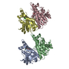
| ||||||||||||||||
|---|---|---|---|---|---|---|---|---|---|---|---|---|---|---|---|---|---|
| 1 | 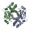
| ||||||||||||||||
| 2 | 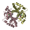
| ||||||||||||||||
| Unit cell |
| ||||||||||||||||
| Atom site foot note | 1: CIS PROLINE - PRO A 56 / 2: CIS PROLINE - PRO B 56 | ||||||||||||||||
| Noncrystallographic symmetry (NCS) | NCS oper:
| ||||||||||||||||
| Details | MTRIX THE TRANSFORMATIONS PRESENTED ON MTRIX RECORDS BELOW DESCRIBE NON-CRYSTALLOGRAPHIC RELATIONSHIPS AMONG THE VARIOUS DOMAINS IN THIS ENTRY. APPLYING THE APPROPRIATE MTRIX TRANSFORMATION TO THE RESIDUES LISTED FIRST WILL YIELD APPROXIMATE COORDINATES FOR THE RESIDUES LISTED SECOND. APPLIED TO TRANSFORMED TO MTRIX RESIDUES RESIDUES RMSD M1 A 2 .. A 438 TRANSFORMS A CHAIN OF DIMER TO A CHAIN OF SECOND DIMER OF THE ASYMMETRIC UNIT M2 B 2 .. B 438 TRANSFORMS B CHAIN OF DIMER TO B CHAIN OF SECOND DIMER OF THE ASYMMETRIC UNIT M3 A 2 .. A 438 B 2 .. B 438 0.001 TRANSFORMS A CHAIN OF DIMER TO B CHAIN OF DIMER |
-Components
| #1: Protein | Mass: 25538.924 Da / Num. of mol.: 4 Source method: isolated from a genetically manipulated source Source: (gene. exp.) Homo sapiens (human) / Organ: LIVER / Plasmid: PTACGST2 / Production host:  #2: Chemical | ChemComp-EAA /   Mass: 303.138 Da / Num. of mol.: 4 / Source method: obtained synthetically / Formula: C13H12Cl2O4 Mass: 303.138 Da / Num. of mol.: 4 / Source method: obtained synthetically / Formula: C13H12Cl2O4#3: Water | ChemComp-HOH / |  Mass: 18.015 Da / Num. of mol.: 150 / Source method: isolated from a natural source / Formula: H2O Mass: 18.015 Da / Num. of mol.: 150 / Source method: isolated from a natural source / Formula: H2OSequence details | RESIDUES ARE NUMBERED FROM 2 TO 222. THE ETHACRYNIC | |
|---|
-Experimental details
-Experiment
| Experiment | Method: X-RAY DIFFRACTION |
|---|
- Sample preparation
| Crystal | Density Matthews: 2.5 Å3/Da / Density % sol: 50.9 % | ||||||||||||||||||||||||||||||||||||||||||||||||||
|---|---|---|---|---|---|---|---|---|---|---|---|---|---|---|---|---|---|---|---|---|---|---|---|---|---|---|---|---|---|---|---|---|---|---|---|---|---|---|---|---|---|---|---|---|---|---|---|---|---|---|---|
| Crystal grow | *PLUS Temperature: 20 ℃ / Method: vapor diffusion | ||||||||||||||||||||||||||||||||||||||||||||||||||
| Components of the solutions | *PLUS
|
-Data collection
| Diffraction source | Wavelength: 1.54 |
|---|---|
| Detector | Type: RIGAKU RAXIS / Detector: IMAGE PLATE / Date: Oct 21, 1993 |
| Radiation | Monochromatic (M) / Laue (L): M / Scattering type: x-ray |
| Radiation wavelength | Wavelength: 1.54 Å / Relative weight: 1 |
| Reflection | Resolution: 2.5→10 Å / Num. obs: 26072 / % possible obs: 94.7 % / Observed criterion σ(I): 0 / Redundancy: 1.6 % / Rmerge(I) obs: 0.078 |
| Reflection | *PLUS Highest resolution: 2.7 Å / Num. measured all: 41606 / Rmerge(I) obs: 0.078 |
- Processing
| Software |
| ||||||||||||||||||||||||||||||||||||||||||||||||||||||||||||
|---|---|---|---|---|---|---|---|---|---|---|---|---|---|---|---|---|---|---|---|---|---|---|---|---|---|---|---|---|---|---|---|---|---|---|---|---|---|---|---|---|---|---|---|---|---|---|---|---|---|---|---|---|---|---|---|---|---|---|---|---|---|
| Refinement | Resolution: 2.7→7.5 Å / σ(F): 0 Details: RESIDUE ARG 89 HAS BEEN MODELLED IN 2 CONFORMATIONS AS FOR THE HIGHER RESOLUTION GST A 1-1 STRUCTURE COMPLEXED WITH A GLUTATHIONE, ETHACRYNIC ACID CONJUGATE. THIS RESIDUE LIES AT THE DIMER ...Details: RESIDUE ARG 89 HAS BEEN MODELLED IN 2 CONFORMATIONS AS FOR THE HIGHER RESOLUTION GST A 1-1 STRUCTURE COMPLEXED WITH A GLUTATHIONE, ETHACRYNIC ACID CONJUGATE. THIS RESIDUE LIES AT THE DIMER INTERFACE CLOSE TO AN NCS COPY OF ITSELF.
| ||||||||||||||||||||||||||||||||||||||||||||||||||||||||||||
| Displacement parameters | Biso mean: 43 Å2 | ||||||||||||||||||||||||||||||||||||||||||||||||||||||||||||
| Refine analyze | Luzzati coordinate error obs: 0.35 Å | ||||||||||||||||||||||||||||||||||||||||||||||||||||||||||||
| Refinement step | Cycle: LAST / Resolution: 2.7→7.5 Å
| ||||||||||||||||||||||||||||||||||||||||||||||||||||||||||||
| Refine LS restraints |
| ||||||||||||||||||||||||||||||||||||||||||||||||||||||||||||
| Software | *PLUS Name: X-PLOR / Classification: refinement | ||||||||||||||||||||||||||||||||||||||||||||||||||||||||||||
| Refine LS restraints | *PLUS
|
