 Movie
Movie Controller

[English] 日本語
 Yorodumi
- PDB-5jcu: Crystal Structure of hGSTA1-1 with Glutathione Adduct of Phenethy... -
+ Open data

- Basic information
| Entry | Database: PDB / ID: 5jcu | ||||||
|---|---|---|---|---|---|---|---|
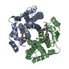
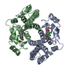
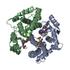
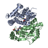
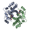
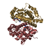
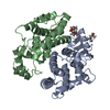
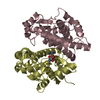
| Title | Crystal Structure of hGSTA1-1 with Glutathione Adduct of Phenethyl Isothiocyanate and Cystein Adduct of Phenethyl Isothiocyanate | ||||||
 Components Components | Glutathione S-transferase A1 | ||||||
 Keywords Keywords | TRANSFERASE / GST / PEITC / Glutathione adduct / Cyctein adduct | ||||||
| Function / homology |  Function and homology information Function and homology informationIsomerases; Intramolecular oxidoreductases; Transposing C=C bonds / glutathione derivative biosynthetic process / steroid Delta-isomerase activity / linoleic acid metabolic process / Glutathione conjugation / glutathione peroxidase activity / Azathioprine ADME / NFE2L2 regulating anti-oxidant/detoxification enzymes / Heme degradation / prostaglandin metabolic process ...Isomerases; Intramolecular oxidoreductases; Transposing C=C bonds / glutathione derivative biosynthetic process / steroid Delta-isomerase activity / linoleic acid metabolic process / Glutathione conjugation / glutathione peroxidase activity / Azathioprine ADME / NFE2L2 regulating anti-oxidant/detoxification enzymes / Heme degradation / prostaglandin metabolic process / glutathione transferase / glutathione transferase activity / Oxidoreductases; Acting on a peroxide as acceptor; Peroxidases / epithelial cell differentiation / xenobiotic metabolic process / fatty acid binding / glutathione metabolic process / extracellular exosome / cytosol Similarity search - Function | ||||||
| Biological species |  Homo sapiens (human) Homo sapiens (human) | ||||||
| Method |  X-RAY DIFFRACTION / SYNCHROTRON / FOURIER SYNTHESIS / Resolution: 1.93 Å X-RAY DIFFRACTION / SYNCHROTRON / FOURIER SYNTHESIS / Resolution: 1.93 Å | ||||||
 Authors Authors | Kumari, V. / Ji, X. | ||||||
 Citation Citation | Journal: Plos One / Year: 2016 Title: Irreversible Inhibition of Glutathione S-Transferase by Phenethyl Isothiocyanate (PEITC), a Dietary Cancer Chemopreventive Phytochemical. Authors: Kumari, V. / Dyba, M.A. / Holland, R.J. / Liang, Y.H. / Singh, S.V. / Ji, X. #1: Journal: J. Mol. Biol. / Year: 1993Title: Structure determination and refinement of human alpha class glutathione transferase A1-1, and a comparison with the Mu and Pi class enzymes. Authors: Sinning, I. / Kleywegt, G.J. / Cowan, S.W. / Reinemer, P. / Dirr, H.W. / Huber, R. / Gilliland, G.L. / Armstrong, R.N. / Ji, X. / Board, P.G. | ||||||
| History |
|
- Structure visualization
| Structure viewer | Molecule: MolmilJmol/JSmol |
|---|
- Downloads & links
-Download
| PDBx/mmCIF format | 5jcu.cif.gz | 219.9 KB | Display | PDBx/mmCIF format |
|---|---|---|---|---|
| PDB format | pdb5jcu.ent.gz | 177.1 KB | Display | PDB format |
| PDBx/mmJSON format | 5jcu.json.gz | Tree view | PDBx/mmJSON format | |
| Others |  Other downloads Other downloads |
-Validation report
| Arichive directory | https://data.pdbj.org/pub/pdb/validation_reports/jc/5jcuftp://data.pdbj.org/pub/pdb/validation_reports/jc/5jcu | HTTPS FTP |
|---|
-Related structure data
| Related structure data | 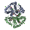 5jcwC  1guhS C: citing same article ( S: Starting model for refinement |
|---|---|
| Similar structure data |


-Links
 PDBj
PDBj


- Assembly
| Deposited unit | 
| ||||||||||||||||||||||||
|---|---|---|---|---|---|---|---|---|---|---|---|---|---|---|---|---|---|---|---|---|---|---|---|---|---|
| 1 | 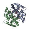
| ||||||||||||||||||||||||
| 2 | 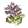
| ||||||||||||||||||||||||
| Unit cell |
| ||||||||||||||||||||||||
| Components on special symmetry positions |
|
-Components
| #1: Protein | Mass: 25702.166 Da / Num. of mol.: 4 Source method: isolated from a genetically manipulated source Source: (gene. exp.) Homo sapiens (human) / Gene: GSTA1 / Plasmid: pET30b(+)/hGSTA1 / Production host:  #2: Chemical | ChemComp-GVX /   Type: peptide-like / Mass: 470.563 Da / Num. of mol.: 4 / Source method: obtained synthetically / Formula: C19H26N4O6S2 Type: peptide-like / Mass: 470.563 Da / Num. of mol.: 4 / Source method: obtained synthetically / Formula: C19H26N4O6S2#3: Chemical | ChemComp-EDO /   Mass: 62.068 Da / Num. of mol.: 6 / Source method: obtained synthetically / Formula: C2H6O2 Mass: 62.068 Da / Num. of mol.: 6 / Source method: obtained synthetically / Formula: C2H6O2#4: Water | ChemComp-HOH / |  Mass: 18.015 Da / Num. of mol.: 1088 / Source method: isolated from a natural source / Formula: H2O Mass: 18.015 Da / Num. of mol.: 1088 / Source method: isolated from a natural source / Formula: H2O |
|---|
-Experimental details
-Experiment
| Experiment | Method: X-RAY DIFFRACTION / Number of used crystals: 1 |
|---|
- Sample preparation
| Crystal | Density Matthews: 2.36 Å3/Da / Density % sol: 47.92 % / Description: Rod |
|---|---|
| Crystal grow | Temperature: 293 K / Method: vapor diffusion, sitting drop / pH: 7.4 / Details: PEG 3350 20% (w/v), Sodium acetate 0.2 M |
-Data collection
| Diffraction | Mean temperature: 100 K |
|---|---|
| Diffraction source | Source: SYNCHROTRON / Site: APS  / Beamline: 22-BM / Wavelength: 1 Å / Beamline: 22-BM / Wavelength: 1 Å |
| Detector | Type: MARMOSAIC 225 mm CCD / Detector: CCD / Date: Nov 29, 2014 / Details: mirrors |
| Radiation | Monochromator: GRAPHITE / Protocol: SINGLE WAVELENGTH / Monochromatic (M) / Laue (L): M / Scattering type: x-ray |
| Radiation wavelength | Wavelength: 1 Å / Relative weight: 1 |
| Reflection | Resolution: 1.93→30 Å / Num. obs: 71288 / % possible obs: 99.2 % / Observed criterion σ(I): -3 / Redundancy: 4.9 % / CC1/2: 0.695 / Rmerge(I) obs: 0.083 / Net I/σ(I): 18.04 |
| Reflection shell | Resolution: 1.93→2 Å / Redundancy: 4.9 % / Rmerge(I) obs: 0.866 / Mean I/σ(I) obs: 1.64 / % possible all: 98.4 |
- Processing
| Software |
| ||||||||||||||||||||||||||||||||||||||||||||||||||||||||
|---|---|---|---|---|---|---|---|---|---|---|---|---|---|---|---|---|---|---|---|---|---|---|---|---|---|---|---|---|---|---|---|---|---|---|---|---|---|---|---|---|---|---|---|---|---|---|---|---|---|---|---|---|---|---|---|---|---|
| Refinement | Method to determine structure: FOURIER SYNTHESIS Starting model: 1GUH Resolution: 1.93→29.799 Å / SU ML: 0.25 / Cross valid method: FREE R-VALUE / σ(F): 1.36 / Phase error: 26.57
| ||||||||||||||||||||||||||||||||||||||||||||||||||||||||
| Solvent computation | Shrinkage radii: 0.9 Å / VDW probe radii: 1.11 Å | ||||||||||||||||||||||||||||||||||||||||||||||||||||||||
| Displacement parameters | Biso mean: 33.2 Å2 | ||||||||||||||||||||||||||||||||||||||||||||||||||||||||
| Refinement step | Cycle: LAST / Resolution: 1.93→29.799 Å
| ||||||||||||||||||||||||||||||||||||||||||||||||||||||||
| Refine LS restraints |
| ||||||||||||||||||||||||||||||||||||||||||||||||||||||||
| LS refinement shell |
|
