 Movie
Movie Controller

[English] 日本語
 Yorodumi
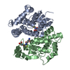
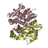
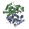
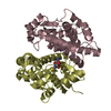
- PDB-1b48: CRYSTAL STRUCTURE OF MGSTA4-4 IN COMPLEX WITH GSH CONJUGATE OF 4-... -
+ Open data

- Basic information
| Entry | Database: PDB / ID: 1b48 | ||||||
|---|---|---|---|---|---|---|---|
| Title | CRYSTAL STRUCTURE OF MGSTA4-4 IN COMPLEX WITH GSH CONJUGATE OF 4-HYDROXYNONENAL IN ONE SUBUNIT AND GSH IN THE OTHER: EVIDENCE OF SIGNALING ACROSS DIMER INTERFACE IN MGSTA4-4 | ||||||
 Components Components | PROTEIN (GLUTATHIONE S-TRANSFERASE) | ||||||
 Keywords Keywords | TRANSFERASE / GLUTATHIONE S-TRANSFERASE / GST / SUBUNIT COOPERATIVITY | ||||||
| Function / homology |  Function and homology information Function and homology informationresponse to caloric restriction / glutathione transferase / glutathione transferase activity / toxic substance binding / xenobiotic metabolic process / glutathione metabolic process / mitochondrion Similarity search - Function | ||||||
| Biological species |  | ||||||
| Method |  X-RAY DIFFRACTION / MOLECULAR REPLACEMENT / Resolution: 2.6 Å X-RAY DIFFRACTION / MOLECULAR REPLACEMENT / Resolution: 2.6 Å | ||||||
 Authors Authors | Xiao, B. / Zimniak, P. / Ji, X. | ||||||
 Citation Citation | Journal: Biochemistry / Year: 1999 Title: Crystal structure of a murine glutathione S-transferase in complex with a glutathione conjugate of 4-hydroxynon-2-enal in one subunit and glutathione in the other: evidence of signaling across the dimer interface. Authors: Xiao, B. / Singh, S.P. / Nanduri, B. / Awasthi, Y.C. / Zimniak, P. / Ji, X. #1: Journal: FEBS Lett. / Year: 1998Title: Crystal Structure of a Murine Alpha-Class Glutathione S-Transferase Involved in Cellular Defense Against Oxidative Stress Authors: Krengel, U. / Schroter, K.H. / Hoier, H. / Arkema, A. / Kalk, K.H. / Zimniak, P. / Dijkstra, B.W. | ||||||
| History |
|
- Structure visualization
| Structure viewer | Molecule: MolmilJmol/JSmol |
|---|
- Downloads & links
-Download
| PDBx/mmCIF format | 1b48.cif.gz | 100.9 KB | Display | PDBx/mmCIF format |
|---|---|---|---|---|
| PDB format | pdb1b48.ent.gz | 78.4 KB | Display | PDB format |
| PDBx/mmJSON format | 1b48.json.gz | Tree view | PDBx/mmJSON format | |
| Others |  Other downloads Other downloads |
-Validation report
| Arichive directory | https://data.pdbj.org/pub/pdb/validation_reports/b4/1b48ftp://data.pdbj.org/pub/pdb/validation_reports/b4/1b48 | HTTPS FTP |
|---|
-Related structure data
| Related structure data |  1guhS S: Starting model for refinement |
|---|---|
| Similar structure data |
-Links
 PDBj
PDBj

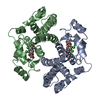
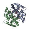
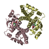
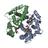
- Assembly
| Deposited unit | 
| ||||||||
|---|---|---|---|---|---|---|---|---|---|
| 1 |
| ||||||||
| Unit cell |
| ||||||||
| Noncrystallographic symmetry (NCS) | NCS oper: (Code: given Matrix: (-0.99956, -0.016641, -0.024543), Vector: |
-Components
| #1: Protein | Mass: 25476.727 Da / Num. of mol.: 2 Source method: isolated from a genetically manipulated source Source: (gene. exp.)  #2: Chemical | ChemComp-HAG / |   Mass: 463.546 Da / Num. of mol.: 1 / Source method: obtained synthetically / Formula: C19H33N3O8S Mass: 463.546 Da / Num. of mol.: 1 / Source method: obtained synthetically / Formula: C19H33N3O8S#3: Chemical | ChemComp-GSH / |   Mass: 307.323 Da / Num. of mol.: 1 / Source method: obtained synthetically / Formula: C10H17N3O6S Mass: 307.323 Da / Num. of mol.: 1 / Source method: obtained synthetically / Formula: C10H17N3O6S#4: Water | ChemComp-HOH / |  Mass: 18.015 Da / Num. of mol.: 40 / Source method: isolated from a natural source / Formula: H2O Mass: 18.015 Da / Num. of mol.: 40 / Source method: isolated from a natural source / Formula: H2O |
|---|
-Experimental details
-Experiment
| Experiment | Method: X-RAY DIFFRACTION / Number of used crystals: 1 |
|---|
- Sample preparation
| Crystal | Density Matthews: 2.35 Å3/Da / Density % sol: 47.6 % | ||||||||||||||||||||||||||||||||||||||||||||||||||||||||||||||||||||||||
|---|---|---|---|---|---|---|---|---|---|---|---|---|---|---|---|---|---|---|---|---|---|---|---|---|---|---|---|---|---|---|---|---|---|---|---|---|---|---|---|---|---|---|---|---|---|---|---|---|---|---|---|---|---|---|---|---|---|---|---|---|---|---|---|---|---|---|---|---|---|---|---|---|---|
| Crystal grow | pH: 7.5 Details: CRYSTALS WERE GROWN IN HANGING DROPS WHICH INITIALLY CONSISTED OF 0.11 MM PROTEIN IN 0.047 M HEPES BUFFER (PH 7.5) CONTAINING 1.82 MM GLUTATHIONE, 10.91 MM HNA-SG (GSH CONJUGATE OF 4- ...Details: CRYSTALS WERE GROWN IN HANGING DROPS WHICH INITIALLY CONSISTED OF 0.11 MM PROTEIN IN 0.047 M HEPES BUFFER (PH 7.5) CONTAINING 1.82 MM GLUTATHIONE, 10.91 MM HNA-SG (GSH CONJUGATE OF 4-HYDROXYNONENAL), 9.1% ETHANOL, 4.5% ISOPROPANOL, AND 6.5% PEG MONOMETHYL ETHER 5K (PH 7.5). THE DROPS WERE EQUILIBRATED AT 293 K AGAINST WELL SOLUTION CONTAINING 10% ISOPROPANOL AND 12% PEG MONOMETHYL ETHER 5K IN 80 MM HEPES BUFFER (PH 7.5). | ||||||||||||||||||||||||||||||||||||||||||||||||||||||||||||||||||||||||
| Crystal | *PLUS | ||||||||||||||||||||||||||||||||||||||||||||||||||||||||||||||||||||||||
| Crystal grow | *PLUS Temperature: 18-20 ℃ / Method: vapor diffusion, hanging drop | ||||||||||||||||||||||||||||||||||||||||||||||||||||||||||||||||||||||||
| Components of the solutions | *PLUS
|
-Data collection
| Diffraction | Mean temperature: 293 K |
|---|---|
| Diffraction source | Source: ROTATING ANODE / Type: ENRAF-NONIUS FR591 / Wavelength: 1.5418 |
| Detector | Type: MAC Science DIP-2020 / Detector: IMAGE PLATE / Date: May 15, 1997 / Details: MIRRORS |
| Radiation | Monochromator: NI FILTER / Protocol: SINGLE WAVELENGTH / Monochromatic (M) / Laue (L): M / Scattering type: x-ray |
| Radiation wavelength | Wavelength: 1.5418 Å / Relative weight: 1 |
| Reflection | Resolution: 2.6→20 Å / Num. obs: 15647 / % possible obs: 87.4 % / Observed criterion σ(I): 0 / Redundancy: 12.1 % / Biso Wilson estimate: 39 Å2 / Rmerge(I) obs: 0.075 / Net I/σ(I): 14 |
| Reflection shell | Resolution: 2.6→2.69 Å / Redundancy: 2.74 % / Rmerge(I) obs: 0.256 / Mean I/σ(I) obs: 2.35 / % possible all: 60.1 |
| Reflection shell | *PLUS % possible obs: 60.1 % |
- Processing
| Software |
| ||||||||||||||||||||||||||||||||||||||||||||||||||||||||||||
|---|---|---|---|---|---|---|---|---|---|---|---|---|---|---|---|---|---|---|---|---|---|---|---|---|---|---|---|---|---|---|---|---|---|---|---|---|---|---|---|---|---|---|---|---|---|---|---|---|---|---|---|---|---|---|---|---|---|---|---|---|---|
| Refinement | Method to determine structure: MOLECULAR REPLACEMENT Starting model: PDB ENTRY 1GUH Resolution: 2.6→20 Å / Data cutoff high absF: 20 / Data cutoff low absF: 2.6 / Cross valid method: FREE R / σ(F): 2 / Details: ALL DATA WERE INCLUDED IN MAP CALCULATIONS.
| ||||||||||||||||||||||||||||||||||||||||||||||||||||||||||||
| Displacement parameters | Biso mean: 49 Å2 | ||||||||||||||||||||||||||||||||||||||||||||||||||||||||||||
| Refinement step | Cycle: LAST / Resolution: 2.6→20 Å
| ||||||||||||||||||||||||||||||||||||||||||||||||||||||||||||
| Refine LS restraints |
| ||||||||||||||||||||||||||||||||||||||||||||||||||||||||||||
| LS refinement shell | Resolution: 2.6→2.72 Å / Total num. of bins used: 8
| ||||||||||||||||||||||||||||||||||||||||||||||||||||||||||||
| Software | *PLUS Name: X-PLOR / Version: 3.843 / Classification: refinement | ||||||||||||||||||||||||||||||||||||||||||||||||||||||||||||
| Refinement | *PLUS σ(F): 2 / % reflection Rfree: 5 % | ||||||||||||||||||||||||||||||||||||||||||||||||||||||||||||
| Solvent computation | *PLUS | ||||||||||||||||||||||||||||||||||||||||||||||||||||||||||||
| Displacement parameters | *PLUS Biso mean: 49 Å2 | ||||||||||||||||||||||||||||||||||||||||||||||||||||||||||||
| Refine LS restraints | *PLUS
|
