 Movie
Movie Controller

[English] 日本語
 Yorodumi
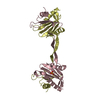
- PDB-1fyk: SERENDIPITOUS CRYSTAL STRUCTURE CONTAINING THE HEAT SHOCK TRANSCR... -
+ Open data

- Basic information
| Entry | Database: PDB / ID: 1fyk | |||||||||
|---|---|---|---|---|---|---|---|---|---|---|
| Title | SERENDIPITOUS CRYSTAL STRUCTURE CONTAINING THE HEAT SHOCK TRANSCRIPTION FACTOR'S DNA BINDING DOMAIN AND COGNATE DNA THAT IS TRANSLATIONALLY DISORDERED | |||||||||
 Components Components |
| |||||||||
 Keywords Keywords | TRANSCRIPTION/DNA / crystal-packing interface / crystallization / protein-DNA interface / protein-protein interface / static disorder / TRANSCRIPTION-DNA COMPLEX | |||||||||
| Function / homology |  Function and homology information Function and homology informationprotein-DNA complex / sequence-specific DNA binding / DNA-binding transcription factor activity / DNA binding / nucleus Similarity search - Function | |||||||||
| Biological species |  Kluyveromyces lactis (yeast) Kluyveromyces lactis (yeast) | |||||||||
| Method |  X-RAY DIFFRACTION / SYNCHROTRON / Resolution: 2.5 Å X-RAY DIFFRACTION / SYNCHROTRON / Resolution: 2.5 Å | |||||||||
 Authors Authors | Littlefield, O. / Nelson, H.C.M. | |||||||||
 Citation Citation | Journal: Proteins / Year: 2001 Title: Crystal packing interaction that blocks crystallization of a site-specific DNA binding protein-DNA complex. Authors: Littlefield, O. / Nelson, H.C. #1: Journal: Nat.Struct.Biol. / Year: 1999Title: A new use for the 'wing' of the 'winged' helix-turn-helix motif in the HSF-DNA cocrystal Authors: Littlefield, O. / Nelson, H.C.M. | |||||||||
| History |
|
- Structure visualization
| Structure viewer | Molecule: MolmilJmol/JSmol |
|---|
- Downloads & links
-Download
| PDBx/mmCIF format | 1fyk.cif.gz | 32.1 KB | Display | PDBx/mmCIF format |
|---|---|---|---|---|
| PDB format | pdb1fyk.ent.gz | 20.9 KB | Display | PDB format |
| PDBx/mmJSON format | 1fyk.json.gz | Tree view | PDBx/mmJSON format | |
| Others |  Other downloads Other downloads |
-Validation report
| Arichive directory | https://data.pdbj.org/pub/pdb/validation_reports/fy/1fykftp://data.pdbj.org/pub/pdb/validation_reports/fy/1fyk | HTTPS FTP |
|---|
-Related structure data


-Links
 PDBj
PDBj- Assembly
| Deposited unit | 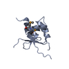
| ||||||||
|---|---|---|---|---|---|---|---|---|---|
| 1 | 
| ||||||||
| Unit cell |
| ||||||||
| Details | protein and DNA are not interacting in a physiologically relevant manner |
-Components
| #1: DNA chain | Mass: 739.422 Da / Num. of mol.: 1 / Source method: obtained synthetically Details: This sequence is based on an idealized HSE sequence. |
|---|---|
| #2: Protein | Mass: 11090.966 Da / Num. of mol.: 1 / Fragment: DNA BINDING DOMAIN / Mutation: N282R, F283H, K284A Source method: isolated from a genetically manipulated source Source: (gene. exp.) Kluyveromyces lactis (yeast) / Plasmid: PHN280R / Production host:  |
| #3: Water | ChemComp-HOH /  Mass: 18.015 Da / Num. of mol.: 38 / Source method: isolated from a natural source / Formula: H2O Mass: 18.015 Da / Num. of mol.: 38 / Source method: isolated from a natural source / Formula: H2O |
| Has protein modification | Y |
-Experimental details
-Experiment
| Experiment | Method: X-RAY DIFFRACTION / Number of used crystals: 1 |
|---|
- Sample preparation
| Crystal | Density Matthews: 2.88 Å3/Da / Density % sol: 57.26 % | ||||||||||||||||||||||||||||||||||||||||||||||||||||||
|---|---|---|---|---|---|---|---|---|---|---|---|---|---|---|---|---|---|---|---|---|---|---|---|---|---|---|---|---|---|---|---|---|---|---|---|---|---|---|---|---|---|---|---|---|---|---|---|---|---|---|---|---|---|---|---|
| Crystal grow | Temperature: 291 K / Method: vapor diffusion, hanging drop / pH: 6 Details: PEG 4000, Cacodylate, Ammonium Acetate, pH 6.0, VAPOR DIFFUSION, HANGING DROP, temperature 291K | ||||||||||||||||||||||||||||||||||||||||||||||||||||||
| Components of the solutions |
| ||||||||||||||||||||||||||||||||||||||||||||||||||||||
| Crystal grow | *PLUS Temperature: 18 ℃ / pH: 7.5 / Method: vapor diffusion | ||||||||||||||||||||||||||||||||||||||||||||||||||||||
| Components of the solutions | *PLUS
|
-Data collection
| Diffraction | Mean temperature: 100 K |
|---|---|
| Diffraction source | Source: SYNCHROTRON / Site: SSRL  / Beamline: BL1-5 / Wavelength: 0.99981 / Beamline: BL1-5 / Wavelength: 0.99981 |
| Detector | Type: FUJI / Detector: IMAGE PLATE / Date: Jan 1, 1995 |
| Radiation | Protocol: SINGLE WAVELENGTH / Monochromatic (M) / Laue (L): M / Scattering type: x-ray |
| Radiation wavelength | Wavelength: 0.99981 Å / Relative weight: 1 |
| Reflection | Resolution: 2.5→20 Å / Num. all: 4940 / Num. obs: 4940 / % possible obs: 98.2 % / Observed criterion σ(F): 0 / Observed criterion σ(I): 0 / Redundancy: 3.7 % / Rmerge(I) obs: 0.05 / Net I/σ(I): 10.3 |
| Reflection shell | Resolution: 2.5→2.55 Å / Redundancy: 3.4 % / Rmerge(I) obs: 0.113 / % possible all: 89.2 |
| Reflection | *PLUS Rmerge(I) obs: 0.05 |
| Reflection shell | *PLUS % possible obs: 89.2 % / Mean I/σ(I) obs: 5.8 |
- Processing
| Software |
| ||||||||||||||||||||
|---|---|---|---|---|---|---|---|---|---|---|---|---|---|---|---|---|---|---|---|---|---|
| Refinement | Resolution: 2.5→20 Å / σ(F): 0 / σ(I): 0 / Stereochemistry target values: Engh & Huber Details: The structure was originally refined by mistake with methionine in lieu of selenomethionine. The deposited coordinates have been adjusted to contain selenomethionine. Because the refinement ...Details: The structure was originally refined by mistake with methionine in lieu of selenomethionine. The deposited coordinates have been adjusted to contain selenomethionine. Because the refinement used group B-factors, the B-factors were kept the same.
| ||||||||||||||||||||
| Refinement step | Cycle: LAST / Resolution: 2.5→20 Å
| ||||||||||||||||||||
| Refine LS restraints |
| ||||||||||||||||||||
| Software | *PLUS Name: X-PLOR / Version: 3.1 / Classification: refinement | ||||||||||||||||||||
| Refinement | *PLUS Highest resolution: 2.5 Å / Lowest resolution: 20 Å / σ(F): 0 / Rfactor obs: 0.209 | ||||||||||||||||||||
| Solvent computation | *PLUS | ||||||||||||||||||||
| Displacement parameters | *PLUS | ||||||||||||||||||||
| LS refinement shell | *PLUS Highest resolution: 2.5 Å / Lowest resolution: 2.59 Å / Rfactor Rfree: 0.358 / Num. reflection Rfree: 54 / Num. reflection Rwork: 419 / Rfactor obs: 0.308 |
