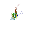
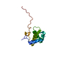
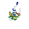
: / PIN domain / 5'-nuclease / Type II/IV secretion system protein / Type II/IV secretion system protein / Large family of predicted nucleotide-binding domains / KH domain / PIN domain / K Homology domain, type 1 / Type-1 KH domain profile. ...: / PIN domain / 5'-nuclease / Type II/IV secretion system protein / Type II/IV secretion system protein / Large family of predicted nucleotide-binding domains / KH domain / PIN domain / K Homology domain, type 1 / Type-1 KH domain profile. / PIN-like domain superfamily / K Homology domain, type 1 superfamily / K Homology domain / K homology RNA-binding domain / K homology domain superfamily, prokaryotic type / Rossmann fold / P-loop containing nucleoside triphosphate hydrolase / 3-Layer(aba) Sandwich / Alpha Beta Similarity search - Domain/homology
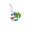
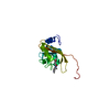
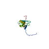
ACETATE ION / FORMIC ACID / DI(HYDROXYETHYL)ETHER / Uncharacterized KH and PIN-domain containing protein MJ1533 Similarity search - Component
Biological species
Methanocaldococcus jannaschii (archaea)
Method
X-RAY DIFFRACTION / SYNCHROTRON / MAD / Resolution: 2.638 Å
In the structure databanks used in Yorodumi, some data are registered as the other names, "COVID-19 virus" and "2019-nCoV". Here are the details of the virus and the list of structure data.
Jan 31, 2019. EMDB accession codes are about to change! (news from PDBe EMDB page)
EMDB accession codes are about to change! (news from PDBe EMDB page)
The allocation of 4 digits for EMDB accession codes will soon come to an end. Whilst these codes will remain in use, new EMDB accession codes will include an additional digit and will expand incrementally as the available range of codes is exhausted. The current 4-digit format prefixed with “EMD-” (i.e. EMD-XXXX) will advance to a 5-digit format (i.e. EMD-XXXXX), and so on. It is currently estimated that the 4-digit codes will be depleted around Spring 2019, at which point the 5-digit format will come into force.
The EM Navigator/Yorodumi systems omit the EMD- prefix.
Related info.:Q: What is EMD? / ID/Accession-code notation in Yorodumi/EM Navigator
Yorodumi is a browser for structure data from EMDB, PDB, SASBDB, etc.
This page is also the successor to EM Navigator detail page, and also detail information page/front-end page for Omokage search.
The word "yorodu" (or yorozu) is an old Japanese word meaning "ten thousand". "mi" (miru) is to see.
Related info.:EMDB / PDB / SASBDB / Comparison of 3 databanks / Yorodumi Search / Aug 31, 2016. New EM Navigator & Yorodumi / Yorodumi Papers / Jmol/JSmol / Function and homology information / Changes in new EM Navigator and Yorodumi
 Movie
Movie Controller

 Yorodumi
 Open data
 Basic information
 Components
 Keywords
 Function and homology information
 Methanocaldococcus jannaschii (archaea)
 X-RAY DIFFRACTION /
  Authors
 Citation
 Structure visualization
 Downloads & links
 Other downloads
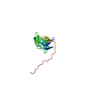
 PDBj
PDBj


 Assembly


 Mass: 35.453 Da / Num. of mol.: 1 / Source method: obtained synthetically / Formula: Cl
 Mass: 106.120 Da / Num. of mol.: 1 / Source method: obtained synthetically / Formula: C4H10O3
 Mass: 46.025 Da / Num. of mol.: 3 / Source method: obtained synthetically / Formula: CH2O2
 Mass: 59.044 Da / Num. of mol.: 2 / Source method: obtained synthetically / Formula: C2H3O2
 Sample preparation
 / Beamline: 19-ID / Wavelength: 0.9794 Å
 Processing