 Movie
Movie Controller


+ Open data

- Basic information
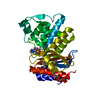
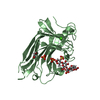
| Entry | Database: PDB / ID: 3hts | ||||||
|---|---|---|---|---|---|---|---|
| Title | HEAT SHOCK TRANSCRIPTION FACTOR/DNA COMPLEX | ||||||
 Components Components |
| ||||||
 Keywords Keywords | TRANSCRIPTION/DNA / TRANSCRIPTION REGULATION / DNA-BINDING PROTEIN / COMPLEX (WINGED HELIX_TURN_ HELIX-DNA) / TRANSCRIPTION-DNA COMPLEX | ||||||
| Function / homology |  Function and homology information Function and homology informationprotein-DNA complex / sequence-specific DNA binding / DNA-binding transcription factor activity / DNA-templated transcription / DNA binding / nucleus Similarity search - Function | ||||||
| Biological species |  Kluyveromyces lactis (yeast) Kluyveromyces lactis (yeast) | ||||||
| Method |  X-RAY DIFFRACTION / SYNCHROTRON / MOLECULAR REPLACEMENT / Resolution: 1.75 Å X-RAY DIFFRACTION / SYNCHROTRON / MOLECULAR REPLACEMENT / Resolution: 1.75 Å | ||||||
 Authors Authors | Littlefield, O. / Nelson, H.C.M. | ||||||
 Citation Citation | Journal: Nat.Struct.Biol. / Year: 1999 Title: A new use for the 'wing' of the 'winged' helix-turn-helix motif in the HSF-DNA cocrystal. Authors: Littlefield, O. / Nelson, H.C. #1: Journal: J.Mol.Biol. / Year: 1995Title: Refinedsolution Structure and Dynamics of the DNA-Binding Domain of the Heat Shock Factor from Kluyveromyces Lactis Authors: Damberger, F.F. / Pelton, J.G. / Liu, C. / Cho, H. / Harrison, C.J. / Nelson, H.C.M. / Wemmer, D.E. #2: Journal: Science / Year: 1994Title: Crystal Structure of the DNA Binding Domain of the Heat Shock Transcription Factor Authors: Harrison, C.J. / Bohm, A.A. / Nelson, H.C.M. | ||||||
| History |
|
- Structure visualization
| Structure viewer | Molecule: MolmilJmol/JSmol |
|---|
- Downloads & links
-Download
| PDBx/mmCIF format | 3hts.cif.gz | 43.4 KB | Display | PDBx/mmCIF format |
|---|---|---|---|---|
| PDB format | pdb3hts.ent.gz | 26.3 KB | Display | PDB format |
| PDBx/mmJSON format | 3hts.json.gz | Tree view | PDBx/mmJSON format | |
| Others |  Other downloads Other downloads |
-Validation report
| Arichive directory | https://data.pdbj.org/pub/pdb/validation_reports/ht/3htsftp://data.pdbj.org/pub/pdb/validation_reports/ht/3hts | HTTPS FTP |
|---|
-Related structure data
| Related structure data |  2htsS S: Starting model for refinement |
|---|---|
| Similar structure data |


-Links
 PDBj
PDBj


- Assembly
| Deposited unit | 
| ||||||||||
|---|---|---|---|---|---|---|---|---|---|---|---|
| 1 | 
| ||||||||||
| Unit cell |
| ||||||||||
| Components on special symmetry positions |
|
-Components
| #1: DNA chain | Mass: 3662.404 Da / Num. of mol.: 1 / Source method: obtained synthetically Details: DESIGN BASED UPON HEAT SHOCK ELEMENT CONSENSUS SEQUENCE |
|---|---|
| #2: Protein | Mass: 11941.409 Da / Num. of mol.: 1 / Fragment: DNA BINDING DOMAIN (RESIDUES 193-281) Source method: isolated from a genetically manipulated source Source: (gene. exp.) Kluyveromyces lactis (yeast) / Species (production host): Escherichia coli / Production host:  |
| #3: Chemical | ChemComp-GOL /   Mass: 92.094 Da / Num. of mol.: 1 / Source method: obtained synthetically / Formula: C3H8O3 Mass: 92.094 Da / Num. of mol.: 1 / Source method: obtained synthetically / Formula: C3H8O3 |
| #4: Water | ChemComp-HOH /  Mass: 18.015 Da / Num. of mol.: 70 / Source method: isolated from a natural source / Formula: H2O Mass: 18.015 Da / Num. of mol.: 70 / Source method: isolated from a natural source / Formula: H2O |
-Experimental details
-Experiment
| Experiment | Method: X-RAY DIFFRACTION / Number of used crystals: 1 |
|---|
- Sample preparation
| Crystal | Density Matthews: 1.89 Å3/Da / Density % sol: 35.02 % Description: LOW RESOLUTION DATA WERE COLLECTED ON A ROTATING ANODE | ||||||||||||||||||||||||||||||||||||||||||
|---|---|---|---|---|---|---|---|---|---|---|---|---|---|---|---|---|---|---|---|---|---|---|---|---|---|---|---|---|---|---|---|---|---|---|---|---|---|---|---|---|---|---|---|
| Crystal grow | pH: 7.5 / Details: pH 7.5 | ||||||||||||||||||||||||||||||||||||||||||
| Crystal grow | *PLUS Method: vapor diffusion | ||||||||||||||||||||||||||||||||||||||||||
| Components of the solutions | *PLUS
|
-Data collection
| Diffraction | Mean temperature: 100 K |
|---|---|
| Diffraction source | Source: SYNCHROTRON / Site: SSRL  / Beamline: BL7-1 / Wavelength: 1.08 / Beamline: BL7-1 / Wavelength: 1.08 |
| Detector | Type: MARRESEARCH / Detector: IMAGE PLATE / Date: May 15, 1996 |
| Radiation | Protocol: SINGLE WAVELENGTH / Monochromatic (M) / Laue (L): M / Scattering type: x-ray |
| Radiation wavelength | Wavelength: 1.08 Å / Relative weight: 1 |
| Reflection | Resolution: 1.75→27.5 Å / Num. obs: 10653 / % possible obs: 89.4 % / Observed criterion σ(I): 1 / Redundancy: 3.2 % / Biso Wilson estimate: 19.9 Å2 / Rmerge(I) obs: 0.035 / Net I/σ(I): 10.3 |
| Reflection shell | Resolution: 1.75→1.8 Å / Redundancy: 2.6 % / Rmerge(I) obs: 0.095 / Mean I/σ(I) obs: 7.4 / % possible all: 86 |
| Reflection | *PLUS Num. measured all: 34273 |
| Reflection shell | *PLUS % possible obs: 86 % / Num. unique obs: 750 / Num. measured obs: 1928 |
- Processing
| Software |
| ||||||||||||||||||||||||||||||||||||||||||||||||||||||||||||||||||||||||||||||||
|---|---|---|---|---|---|---|---|---|---|---|---|---|---|---|---|---|---|---|---|---|---|---|---|---|---|---|---|---|---|---|---|---|---|---|---|---|---|---|---|---|---|---|---|---|---|---|---|---|---|---|---|---|---|---|---|---|---|---|---|---|---|---|---|---|---|---|---|---|---|---|---|---|---|---|---|---|---|---|---|---|---|
| Refinement | Method to determine structure: MOLECULAR REPLACEMENT Starting model: 2HTS Resolution: 1.75→20 Å / Rfactor Rfree error: 0.007 / Data cutoff high absF: 10000000 / Data cutoff low absF: 0.0001 / Isotropic thermal model: RESTRAINED / Cross valid method: THROUGHOUT / σ(F): 0 / Details: BULK SOLVENT MODEL USED
| ||||||||||||||||||||||||||||||||||||||||||||||||||||||||||||||||||||||||||||||||
| Displacement parameters | Biso mean: 38.5 Å2
| ||||||||||||||||||||||||||||||||||||||||||||||||||||||||||||||||||||||||||||||||
| Refine analyze |
| ||||||||||||||||||||||||||||||||||||||||||||||||||||||||||||||||||||||||||||||||
| Refinement step | Cycle: LAST / Resolution: 1.75→20 Å
| ||||||||||||||||||||||||||||||||||||||||||||||||||||||||||||||||||||||||||||||||
| Refine LS restraints |
| ||||||||||||||||||||||||||||||||||||||||||||||||||||||||||||||||||||||||||||||||
| LS refinement shell | Resolution: 1.75→1.86 Å / Rfactor Rfree error: 0.025 / Total num. of bins used: 6
| ||||||||||||||||||||||||||||||||||||||||||||||||||||||||||||||||||||||||||||||||
| Xplor file |
| ||||||||||||||||||||||||||||||||||||||||||||||||||||||||||||||||||||||||||||||||
| Software | *PLUS Name: X-PLOR / Version: 3.851 / Classification: refinement | ||||||||||||||||||||||||||||||||||||||||||||||||||||||||||||||||||||||||||||||||
| Refinement | *PLUS Lowest resolution: 20 Å / σ(F): 0 / % reflection Rfree: 10.5 % | ||||||||||||||||||||||||||||||||||||||||||||||||||||||||||||||||||||||||||||||||
| Solvent computation | *PLUS | ||||||||||||||||||||||||||||||||||||||||||||||||||||||||||||||||||||||||||||||||
| Displacement parameters | *PLUS Biso mean: 38.5 Å2 | ||||||||||||||||||||||||||||||||||||||||||||||||||||||||||||||||||||||||||||||||
| Refine LS restraints | *PLUS
| ||||||||||||||||||||||||||||||||||||||||||||||||||||||||||||||||||||||||||||||||
| LS refinement shell | *PLUS Rfactor Rfree: 0.319 / % reflection Rfree: 9.5 % / Rfactor Rwork: 0.323 / Rfactor obs: 0.323 |
