HETEROGEN Apparent short contacts between the 2 intermediates, TBE and MDD, arise because the ...HETEROGEN Apparent short contacts between the 2 intermediates, TBE and MDD, arise because the crystal structure contains a composite of 2 complexes, one a breakdown product of the other.
Resolution: 1.8→20 Å / Cross valid method: THROUGHOUT / σ(F): 0 / σ(I): 0 / Stereochemistry target values: Engh & Huber Details: Atoms having alternate conformations are only listed once in the above list of the number of atoms used in the refinement
Rfactor
Num. reflection
% reflection
Selection details
Rfree
0.176
964
-
random
Rwork
0.14
-
-
-
all
0.141
22020
-
-
obs
0.141
20094
91.3 %
-
Displacement parameters
Biso mean: 10.9 Å2
Refine analyze
Free
Obs
Luzzati coordinate error
0.18 Å
0.14 Å
Luzzati d res low
-
5 Å
Luzzati sigma a
0.13 Å
0.11 Å
Refinement step
Cycle: LAST / Resolution: 1.8→20 Å
Protein
Nucleic acid
Ligand
Solvent
Total
Num. atoms
2022
0
86
396
2504
Refine LS restraints
Refine-ID
Type
Dev ideal
X-RAY DIFFRACTION
c_bond_d
0.007
X-RAY DIFFRACTION
c_angle_deg
1.4
X-RAY DIFFRACTION
c_dihedral_angle_d
24.5
X-RAY DIFFRACTION
c_improper_angle_d
0.8
LS refinement shell
Resolution (Å)
Rfactor Rfree
Num. reflection Rfree
Rfactor Rwork
Refine-ID
Num. reflection obs
% reflection obs (%)
1.8-1.91
0.2036
151
0.1753
X-RAY DIFFRACTION
2769
79
1.91-2.06
0.1973
145
0.1487
X-RAY DIFFRACTION
3147
89
2.06-2.27
0.187
143
0.1462
X-RAY DIFFRACTION
3309
92
2.27-2.6
0.1808
166
0.1336
X-RAY DIFFRACTION
3433
95
2.6-3.27
0.1753
176
0.1368
X-RAY DIFFRACTION
3608
98
3.27-20
0.1511
183
0.1291
X-RAY DIFFRACTION
3828
100
+
About Yorodumi
-
News
-
Feb 9, 2022. New format data for meta-information of EMDB entries
New format data for meta-information of EMDB entries
Version 3 of the EMDB header file is now the official format.
The previous official version 1.9 will be removed from the archive.
In the structure databanks used in Yorodumi, some data are registered as the other names, "COVID-19 virus" and "2019-nCoV". Here are the details of the virus and the list of structure data.
Jan 31, 2019. EMDB accession codes are about to change! (news from PDBe EMDB page)
EMDB accession codes are about to change! (news from PDBe EMDB page)
The allocation of 4 digits for EMDB accession codes will soon come to an end. Whilst these codes will remain in use, new EMDB accession codes will include an additional digit and will expand incrementally as the available range of codes is exhausted. The current 4-digit format prefixed with “EMD-” (i.e. EMD-XXXX) will advance to a 5-digit format (i.e. EMD-XXXXX), and so on. It is currently estimated that the 4-digit codes will be depleted around Spring 2019, at which point the 5-digit format will come into force.
The EM Navigator/Yorodumi systems omit the EMD- prefix.
Related info.:Q: What is EMD? / ID/Accession-code notation in Yorodumi/EM Navigator
Yorodumi is a browser for structure data from EMDB, PDB, SASBDB, etc.
This page is also the successor to EM Navigator detail page, and also detail information page/front-end page for Omokage search.
The word "yorodu" (or yorozu) is an old Japanese word meaning "ten thousand". "mi" (miru) is to see.
Related info.:EMDB / PDB / SASBDB / Comparison of 3 databanks / Yorodumi Search / Aug 31, 2016. New EM Navigator & Yorodumi / Yorodumi Papers / Jmol/JSmol / Function and homology information / Changes in new EM Navigator and Yorodumi
 Movie
Movie Controller


 Open data
 Basic information
 Components
 Keywords
 Function and homology information
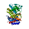
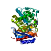
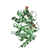
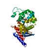
 Klebsiella pneumoniae (bacteria)
 X-RAY DIFFRACTION /
  Authors
 Citation
 Structure visualization
 Downloads & links
 Other downloads


 PDBj
PDBj


 Assembly
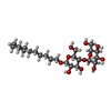


 Mass: 508.600 Da / Num. of mol.: 2 / Source method: obtained synthetically / Formula: C24H44O11
 Mass: 302.307 Da / Num. of mol.: 1 / Source method: obtained synthetically / Formula: C10H14N4O5S
 Mass: 72.063 Da / Num. of mol.: 1 / Source method: obtained synthetically / Formula: C3H4O2
 Mass: 118.174 Da / Num. of mol.: 1 / Source method: obtained synthetically / Formula: C6H14O2 / Comment: precipitant*YM
 Mass: 118.174 Da / Num. of mol.: 1 / Source method: obtained synthetically / Formula: C6H14O2 / Comment: precipitant*YM
 Sample preparation
 Processing