 Movie
Movie Controller


+ Open data

- Basic information
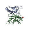
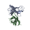
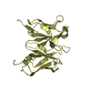
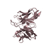
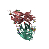
| Entry | Database: PDB / ID: 1eeu | ||||||
|---|---|---|---|---|---|---|---|
| Title | M4L/Y(27D)D/Q89D/T94H mutant of LEN | ||||||
 Components Components | KAPPA-4 IMMUNOGLOBULIN (LIGHT CHAIN) | ||||||
 Keywords Keywords |  IMMUNE SYSTEM / Human Kappa-4 Immunoglobulin Light Chain / Mutant / Aspartic Acid in beta-sheet / Protein Stability IMMUNE SYSTEM / Human Kappa-4 Immunoglobulin Light Chain / Mutant / Aspartic Acid in beta-sheet / Protein Stability | ||||||
| Function / homology |  Function and homology information Function and homology informationCD22 mediated BCR regulation / Fc epsilon receptor (FCERI) signaling / Classical antibody-mediated complement activation / Initial triggering of complement / immunoglobulin complex / FCGR activation / Role of phospholipids in phagocytosis / Role of LAT2/NTAL/LAB on calcium mobilization / Scavenging of heme from plasma / antigen binding ...CD22 mediated BCR regulation / Fc epsilon receptor (FCERI) signaling / Classical antibody-mediated complement activation / Initial triggering of complement / immunoglobulin complex / FCGR activation / Role of phospholipids in phagocytosis / Role of LAT2/NTAL/LAB on calcium mobilization / Scavenging of heme from plasma / antigen binding / FCERI mediated Ca+2 mobilization / FCGR3A-mediated IL10 synthesis / Antigen activates B Cell Receptor (BCR) leading to generation of second messengers / Regulation of Complement cascade / Cell surface interactions at the vascular wall / FCGR3A-mediated phagocytosis / FCERI mediated MAPK activation / Regulation of actin dynamics for phagocytic cup formation / FCERI mediated NF-kB activation / Immunoregulatory interactions between a Lymphoid and a non-Lymphoid cell / blood microparticle / Potential therapeutics for SARS / adaptive immune response / immune response / extracellular space / extracellular region / plasma membraneSimilarity search - Function | ||||||
| Biological species |  Homo sapiens (human) Homo sapiens (human) | ||||||
| Method | X-RAY DIFFRACTION / SYNCHROTRON / Rigid body fitting / Resolution: 1.6 Å | ||||||
 Authors Authors | Pokkuluri, P.R. / Cai, X. / Gu, M. / Stevens, F.J. / Schiffer, M. | ||||||
 Citation Citation | Journal: Protein Sci. / Year: 2002 Title: Factors contributing to decreased protein stability when aspartic acid residues are in beta-sheet regions. Authors: Pokkuluri, P.R. / Gu, M. / Cai, X. / Raffen, R. / Stevens, F.J. / Schiffer, M. | ||||||
| History |
|
- Structure visualization
| Structure viewer | Molecule: MolmilJmol/JSmol |
|---|
- Downloads & links
-Download
| PDBx/mmCIF format | 1eeu.cif.gz | 64.4 KB | Display | PDBx/mmCIF format |
|---|---|---|---|---|
| PDB format | pdb1eeu.ent.gz | 45.7 KB | Display | PDB format |
| PDBx/mmJSON format | 1eeu.json.gz | Tree view | PDBx/mmJSON format | |
| Others |  Other downloads Other downloads |
-Validation report
| Arichive directory | https://data.pdbj.org/pub/pdb/validation_reports/ee/1eeuftp://data.pdbj.org/pub/pdb/validation_reports/ee/1eeu | HTTPS FTP |
|---|
-Related structure data
| Related structure data |  1efqC  1eeqS S: Starting model for refinement C: citing same article ( |
|---|---|
| Similar structure data |

-Links
 PDBj
PDBj


- Assembly
| Deposited unit | 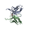
| ||||||||
|---|---|---|---|---|---|---|---|---|---|
| 1 |
| ||||||||
| Unit cell |
| ||||||||
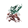
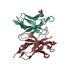
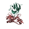
| Details | The biological Assembly is a homo dimer formed by chains A and B. |
-Components
| #1: Antibody | Mass: 12607.890 Da / Num. of mol.: 2 / Mutation: M4L/Y(27D)D/Q89D/T94H Source method: isolated from a genetically manipulated source Source: (gene. exp.) Homo sapiens (human) / Plasmid: PASK40 / Production host:  Escherichia coli (E. coli) / References: UniProt: P01625, UniProt: P06312*PLUS Escherichia coli (E. coli) / References: UniProt: P01625, UniProt: P06312*PLUS#2: Chemical | Isopropyl alcohol  Mass: 60.095 Da / Num. of mol.: 2 / Source method: obtained synthetically / Formula: C3H8O / Comment: alkaloid*YM Mass: 60.095 Da / Num. of mol.: 2 / Source method: obtained synthetically / Formula: C3H8O / Comment: alkaloid*YM#3: Water | ChemComp-HOH / | Water Mass: 18.015 Da / Num. of mol.: 334 / Source method: isolated from a natural source / Formula: H2O Mass: 18.015 Da / Num. of mol.: 334 / Source method: isolated from a natural source / Formula: H2O |
|---|
-Experimental details
-Experiment
| Experiment | Method: X-RAY DIFFRACTION / Number of used crystals: 1 |
|---|
- Sample preparation
| Crystal | Density Matthews: 2.77 Å3/Da / Density % sol: 55.61 % | ||||||||||||||||||||||||||||||||||||
|---|---|---|---|---|---|---|---|---|---|---|---|---|---|---|---|---|---|---|---|---|---|---|---|---|---|---|---|---|---|---|---|---|---|---|---|---|---|
| Crystal grow | Temperature: 298 K / Method: vapor diffusion, hanging drop / pH: 5.6 Details: 20% PEG4K, 20% 2-Propanol, 0.1 M Sodium Citrate pH 5.6, VAPOR DIFFUSION, HANGING DROP, temperature 298K | ||||||||||||||||||||||||||||||||||||
| Crystal grow | *PLUS | ||||||||||||||||||||||||||||||||||||
| Components of the solutions | *PLUS
|
-Data collection
| Diffraction | Mean temperature: 100 K |
|---|---|
| Diffraction source | Source: SYNCHROTRON / Site: APS  / Beamline: 19-ID / Wavelength: 0.91841 / Beamline: 19-ID / Wavelength: 0.91841 |
| Detector | Type: CUSTOM-MADE / Detector: CCD / Date: Jul 6, 1999 |
| Radiation | Protocol: SINGLE WAVELENGTH / Monochromatic (M) / Laue (L): M / Scattering type: x-ray |
| Radiation wavelength | Wavelength: 0.91841 Å / Relative weight: 1 |
| Reflection | Resolution: 1.6→20 Å / Num. all: 37837 / Num. obs: 37705 / % possible obs: 99.7 % / Observed criterion σ(I): -3 / Redundancy: 18 % / Biso Wilson estimate: 17 Å2 / Rmerge(I) obs: 0.061 / Net I/σ(I): 43.6 |
| Reflection shell | Resolution: 1.6→1.65 Å / Redundancy: 6 % / Rmerge(I) obs: 0.26 / Mean I/σ(I) obs: 7.3 / Num. unique all: 3491 / % possible all: 99.6 |
| Reflection shell | *PLUS Rmerge(I) obs: 0.263 |
- Processing
| Software |
| ||||||||||||||||||||||||||||||||||||||||
|---|---|---|---|---|---|---|---|---|---|---|---|---|---|---|---|---|---|---|---|---|---|---|---|---|---|---|---|---|---|---|---|---|---|---|---|---|---|---|---|---|---|
| Refinement | Method to determine structure: Rigid body fitting Starting model: 1EEQ WITH ATOMS BEYOND CB REMOVED FOR RESIDUES A 89 AND B 389 Resolution: 1.6→8 Å / Rfactor Rfree error: 0.004 / Data cutoff high absF: 763311.74 / Data cutoff low absF: 0 / Isotropic thermal model: RESTRAINED / Cross valid method: THROUGHOUT / σ(F): 3 / Stereochemistry target values: Engh & Huber
| ||||||||||||||||||||||||||||||||||||||||
| Solvent computation | Solvent model: FLAT MODEL / Bsol: 62.31 Å2 / ksol: 0.515 e/Å3 | ||||||||||||||||||||||||||||||||||||||||
| Displacement parameters | Biso mean: 20.1 Å2
| ||||||||||||||||||||||||||||||||||||||||
| Refine analyze |
| ||||||||||||||||||||||||||||||||||||||||
| Refinement step | Cycle: LAST / Resolution: 1.6→8 Å
| ||||||||||||||||||||||||||||||||||||||||
| Refine LS restraints |
| ||||||||||||||||||||||||||||||||||||||||
| LS refinement shell | Resolution: 1.6→1.7 Å / Rfactor Rfree error: 0.011 / Total num. of bins used: 6
| ||||||||||||||||||||||||||||||||||||||||
| Xplor file |
| ||||||||||||||||||||||||||||||||||||||||
| Software | *PLUS Name: CNS / Version: 0.9 / Classification: refinement | ||||||||||||||||||||||||||||||||||||||||
| Refinement | *PLUS σ(F): 3 / % reflection Rfree: 10 % / Rfactor obs: 0.195 | ||||||||||||||||||||||||||||||||||||||||
| Solvent computation | *PLUS | ||||||||||||||||||||||||||||||||||||||||
| Displacement parameters | *PLUS Biso mean: 20.1 Å2 | ||||||||||||||||||||||||||||||||||||||||
| Refine LS restraints | *PLUS
| ||||||||||||||||||||||||||||||||||||||||
| LS refinement shell | *PLUS Rfactor Rfree: 0.236 / % reflection Rfree: 10.2 % / Rfactor Rwork: 0.204 |
