 Movie
Movie Controller

[English] 日本語
 Yorodumi
- PDB-1dot: CRYSTALLOGRAPHIC STRUCTURE OF DUCK OVOTRANSFERRIN AT 2.3 ANGSTROM... -
+ Open data

- Basic information
| Entry | Database: PDB / ID: 1dot | ||||||
|---|---|---|---|---|---|---|---|
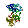
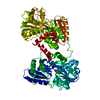
| Title | CRYSTALLOGRAPHIC STRUCTURE OF DUCK OVOTRANSFERRIN AT 2.3 ANGSTROMS RESOLUTION | ||||||
 Components Components | DUCK OVOTRANSFERRIN | ||||||
 Keywords Keywords | IRON TRANSPORT PROTEIN | ||||||
| Function / homology |  Function and homology information Function and homology informationiron ion transport / recycling endosome / antibacterial humoral response / early endosome / extracellular space / metal ion binding / plasma membrane Similarity search - Function | ||||||
| Biological species |  | ||||||
| Method |  X-RAY DIFFRACTION / SYNCHROTRON / Resolution: 2.35 Å X-RAY DIFFRACTION / SYNCHROTRON / Resolution: 2.35 Å | ||||||
 Authors Authors | Rawas, A. / Muirhead, H. | ||||||
 Citation Citation | Journal: Acta Crystallogr.,Sect.D / Year: 1996 Title: Structure of diferric duck ovotransferrin at 2.35 A resolution. Authors: Rawas, A. / Muirhead, H. / Williams, J. #1: Journal: J.Mol.Biol. / Year: 1989Title: Preliminary Crystallographic Studies on Duck Ovotransferrin Authors: Rawas, A. / Moreton, K. / Muirhead, H. / Williams, J. #2: Journal: Biochemistry / Year: 1988Title: Molecular Structure of Serum Transferrin at 3.3 Angstroms Resolution Authors: Bailey, S. / Evans, R.W. / Garratt, R.C. / Gorinsky, B. / Hasnain, S. / Horsburgh, C. / Jhoti, H. / Lindley, P.F. / Mydin, A. / Sarra, R. / Watson, J.L. #3: Journal: Proc.Natl.Acad.Sci.USA / Year: 1987Title: Structure of Human Lactoferrin at 3.2 Angstroms Resolution Authors: Anderson, B.F. / Baker, H.M. / Dodson, E.J. / Norris, G.E. / Rumball, S.V. / Waters, J.M. / Baker, E.N. | ||||||
| History |
| ||||||
| Remark 700 | SHEET SHEETS N1, N2, C1, AND C2 ARE MIXED BETA SHEETS. |
- Structure visualization
| Structure viewer | Molecule: MolmilJmol/JSmol |
|---|
- Downloads & links
-Download
| PDBx/mmCIF format | 1dot.cif.gz | 160.2 KB | Display | PDBx/mmCIF format |
|---|---|---|---|---|
| PDB format | pdb1dot.ent.gz | 118.9 KB | Display | PDB format |
| PDBx/mmJSON format | 1dot.json.gz | Tree view | PDBx/mmJSON format | |
| Others |  Other downloads Other downloads |
-Validation report
| Arichive directory | https://data.pdbj.org/pub/pdb/validation_reports/do/1dotftp://data.pdbj.org/pub/pdb/validation_reports/do/1dot | HTTPS FTP |
|---|
-Related structure data
| Similar structure data |
|---|
-Links
 PDBj
PDBj


- Assembly
| Deposited unit | 
| ||||||||
|---|---|---|---|---|---|---|---|---|---|
| 1 |
| ||||||||
| Unit cell |
| ||||||||
| Atom site foot note | 1: PRO 2 - PRO 3 OMEGA = 149.33 PEPTIDE BOND DEVIATES SIGNIFICANTLY FROM TRANS CONFORMATION 2: PRO 3 - LYS 4 OMEGA = 132.22 PEPTIDE BOND DEVIATES SIGNIFICANTLY FROM TRANS CONFORMATION 3: CIS PROLINE - PRO 71 4: THR 279 - SER 280 OMEGA = 300.70 PEPTIDE BOND DEVIATES SIGNIFICANTLY FROM TRANS CONFORMATION 5: LYS 344 - ILE 345 OMEGA = 127.92 PEPTIDE BOND DEVIATES SIGNIFICANTLY FROM TRANS CONFORMATION |
-Components
-Protein , 1 types, 1 molecules A
| #1: Protein | Mass: 75731.805 Da / Num. of mol.: 1 / Source method: isolated from a natural source / Source: (natural) |
|---|
-Sugars , 2 types, 3 molecules 

| #2: Sugar |  Type: D-saccharide, beta linking / Mass: 221.208 Da / Num. of mol.: 2 Type: D-saccharide, beta linking / Mass: 221.208 Da / Num. of mol.: 2Source method: isolated from a genetically manipulated source Formula: C8H15NO6 #3: Sugar | ChemComp-FUC / |  Type: L-saccharide, alpha linking / Mass: 164.156 Da / Num. of mol.: 1 Type: L-saccharide, alpha linking / Mass: 164.156 Da / Num. of mol.: 1Source method: isolated from a genetically manipulated source Formula: C6H12O5 |
|---|
-Non-polymers , 3 types, 322 molecules 


| #4: Chemical |  Mass: 55.845 Da / Num. of mol.: 2 / Source method: obtained synthetically / Formula: Fe Mass: 55.845 Da / Num. of mol.: 2 / Source method: obtained synthetically / Formula: Fe#5: Chemical |  Mass: 60.009 Da / Num. of mol.: 2 / Source method: obtained synthetically / Formula: CO3 Mass: 60.009 Da / Num. of mol.: 2 / Source method: obtained synthetically / Formula: CO3#6: Water | ChemComp-HOH / | Mass: 18.015 Da / Num. of mol.: 318 / Source method: isolated from a natural source / Formula: H2O |
|---|
-Details
| Has protein modification | Y |
|---|
-Experimental details
-Experiment
| Experiment | Method: X-RAY DIFFRACTION |
|---|
- Sample preparation
| Crystal | Density Matthews: 2.5 Å3/Da / Density % sol: 50.88 % | ||||||||||||||||||||
|---|---|---|---|---|---|---|---|---|---|---|---|---|---|---|---|---|---|---|---|---|---|
| Crystal | *PLUS Density % sol: 48 % | ||||||||||||||||||||
| Crystal grow | *PLUS Temperature: 291 K / pH: 5.8 / Method: batch method | ||||||||||||||||||||
| Components of the solutions | *PLUS
|
-Data collection
| Diffraction source | Source: SYNCHROTRON / Site: SRS  / Beamline: PX7.2 / Wavelength: 1.488 / Beamline: PX7.2 / Wavelength: 1.488 |
|---|---|
| Detector | Detector: FILM / Date: 1986 |
| Radiation | Monochromatic (M) / Laue (L): M / Scattering type: x-ray |
| Radiation wavelength | Wavelength: 1.488 Å / Relative weight: 1 |
| Reflection | Num. obs: 29846 / % possible obs: 90 % / Rmerge(I) obs: 0.059 |
| Reflection | *PLUS Highest resolution: 2.35 Å / Lowest resolution: 10 Å / Rmerge(I) obs: 0.059 |
- Processing
| Software |
| ||||||||||||||||||||||||||||||||||||||||||||||||||||||||||||||||||||||||||||||||
|---|---|---|---|---|---|---|---|---|---|---|---|---|---|---|---|---|---|---|---|---|---|---|---|---|---|---|---|---|---|---|---|---|---|---|---|---|---|---|---|---|---|---|---|---|---|---|---|---|---|---|---|---|---|---|---|---|---|---|---|---|---|---|---|---|---|---|---|---|---|---|---|---|---|---|---|---|---|---|---|---|---|
| Refinement | Resolution: 2.35→10 Å / σ(F): 1
| ||||||||||||||||||||||||||||||||||||||||||||||||||||||||||||||||||||||||||||||||
| Displacement parameters | Biso mean: 28.7 Å2 | ||||||||||||||||||||||||||||||||||||||||||||||||||||||||||||||||||||||||||||||||
| Refinement step | Cycle: LAST / Resolution: 2.35→10 Å
| ||||||||||||||||||||||||||||||||||||||||||||||||||||||||||||||||||||||||||||||||
| Refine LS restraints |
| ||||||||||||||||||||||||||||||||||||||||||||||||||||||||||||||||||||||||||||||||
| Software | *PLUS Name: X-PLOR / Classification: refinement | ||||||||||||||||||||||||||||||||||||||||||||||||||||||||||||||||||||||||||||||||
| Refinement | *PLUS | ||||||||||||||||||||||||||||||||||||||||||||||||||||||||||||||||||||||||||||||||
| Solvent computation | *PLUS | ||||||||||||||||||||||||||||||||||||||||||||||||||||||||||||||||||||||||||||||||
| Displacement parameters | *PLUS | ||||||||||||||||||||||||||||||||||||||||||||||||||||||||||||||||||||||||||||||||
| Refine LS restraints | *PLUS
|
