 Movie
Movie Controller

[English] 日本語
 Yorodumi
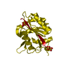
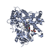
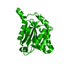
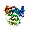
- PDB-1di7: 1.60 ANGSTROM CRYSTAL STRUCTURE OF THE MOLYBDENUM COFACTOR BIOSYN... -
+ Open data

- Basic information
| Entry | Database: PDB / ID: 1di7 | ||||||
|---|---|---|---|---|---|---|---|
| Title | 1.60 ANGSTROM CRYSTAL STRUCTURE OF THE MOLYBDENUM COFACTOR BIOSYNTHESIS PROTEIN MOGA FROM ESCHERICHIA COLI | ||||||
 Components Components | MOLYBDENUM COFACTOR BIOSYNTHETIC ENZYME | ||||||
 Keywords Keywords | UNKNOWN FUNCTION /  MOLYBDENUM COFACTOR / MOCO / MOYBDENUM CO-FACTOR / MOG / MOGA / GEPHYRIN MOLYBDENUM COFACTOR / MOCO / MOYBDENUM CO-FACTOR / MOG / MOGA / GEPHYRIN | ||||||
| Function / homology |  Function and homology information Function and homology informationmolybdopterin cofactor biosynthetic process / molybdopterin adenylyltransferase / molybdopterin adenylyltransferase activity / Mo-molybdopterin cofactor biosynthetic process / ATP binding / identical protein binding / cytosolSimilarity search - Function | ||||||
| Biological species |  Escherichia coli (E. coli) Escherichia coli (E. coli) | ||||||
| Method | X-RAY DIFFRACTION / SYNCHROTRON / MIR / Resolution: 1.6 Å | ||||||
 Authors Authors | Liu, M.T.W. / Wuebbens, M.M. / Rajagopalan, K.V. / Schindelin, H. | ||||||
 Citation Citation | Journal: J.Biol.Chem. / Year: 2000 Title: Crystal structure of the gephyrin-related molybdenum cofactor biosynthesis protein MogA from Escherichia coli. Authors: Liu, M.T. / Wuebbens, M.M. / Rajagopalan, K.V. / Schindelin, H. | ||||||
| History |
|
- Structure visualization
| Structure viewer | Molecule: MolmilJmol/JSmol |
|---|
- Downloads & links
-Download
| PDBx/mmCIF format | 1di7.cif.gz | 51.4 KB | Display | PDBx/mmCIF format |
|---|---|---|---|---|
| PDB format | pdb1di7.ent.gz | 36.6 KB | Display | PDB format |
| PDBx/mmJSON format | 1di7.json.gz | Tree view | PDBx/mmJSON format | |
| Others |  Other downloads Other downloads |
-Validation report
| Arichive directory | https://data.pdbj.org/pub/pdb/validation_reports/di/1di7ftp://data.pdbj.org/pub/pdb/validation_reports/di/1di7 | HTTPS FTP |
|---|
-Related structure data


-Links
 PDBj
PDBj- Assembly
| Deposited unit | 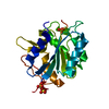
| ||||||||
|---|---|---|---|---|---|---|---|---|---|
| 1 | 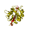
| ||||||||
| Unit cell |
| ||||||||
| Components on special symmetry positions |
|
-Components
| #1: Protein | Mass: 21200.273 Da / Num. of mol.: 1 / Mutation: N2A Source method: isolated from a genetically manipulated source Source: (gene. exp.) Escherichia coli (E. coli) / Production host: Escherichia coli (E. coli) / References: UniProt: P28694, UniProt: P0AF03*PLUS | ||
|---|---|---|---|
| #2: Chemical | Sulfate  Mass: 96.063 Da / Num. of mol.: 3 / Source method: obtained synthetically / Formula: SO4 Mass: 96.063 Da / Num. of mol.: 3 / Source method: obtained synthetically / Formula: SO4#3: Water | ChemComp-HOH / | Water Mass: 18.015 Da / Num. of mol.: 160 / Source method: isolated from a natural source / Formula: H2O Mass: 18.015 Da / Num. of mol.: 160 / Source method: isolated from a natural source / Formula: H2O |
-Experimental details
-Experiment
| Experiment | Method: X-RAY DIFFRACTION / Number of used crystals: 1 |
|---|
- Sample preparation
| Crystal | Density Matthews: 1.9 Å3/Da / Density % sol: 34 % | |||||||||||||||
|---|---|---|---|---|---|---|---|---|---|---|---|---|---|---|---|---|
| Crystal grow | Temperature: 295 K / Method: vapor diffusion / pH: 9 Details: 2.3 - 2.4 M AMMONIUM SULFATE, 0.1 M BICINE pH 9.0, VAPOR DIFFUSION, temperature 295K | |||||||||||||||
| Crystal grow | *PLUS pH: 7.5 | |||||||||||||||
| Components of the solutions | *PLUS
|
-Data collection
| Diffraction | Mean temperature: 95 K |
|---|---|
| Diffraction source | Source: SYNCHROTRON / Site: NSLS  / Beamline: X26C / Wavelength: 1.1 / Beamline: X26C / Wavelength: 1.1 |
| Detector | Type: MARRESEARCH / Detector: IMAGE PLATE / Date: Oct 8, 1998 |
| Radiation | Protocol: SINGLE WAVELENGTH / Monochromatic (M) / Laue (L): M / Scattering type: x-ray |
| Radiation wavelength | Wavelength: 1.1 Å / Relative weight: 1 |
| Reflection | Resolution: 1.6→50 Å / Num. all: 83186 / Num. obs: 18480 / % possible obs: 91.5 % / Observed criterion σ(F): 0 / Observed criterion σ(I): 0 / Redundancy: 4.4 % / Biso Wilson estimate: 15 Å2 / Rmerge(I) obs: 0.052 / Net I/σ(I): 18 |
| Reflection shell | Resolution: 1.6→1.66 Å / Rmerge(I) obs: 0.289 / Mean I/σ(I) obs: 2.7 / % possible all: 97.7 |
| Reflection shell | *PLUS % possible obs: 97.7 % |
- Processing
| Software |
| ||||||||||||||||||||||||||||||||||||||||||||||||
|---|---|---|---|---|---|---|---|---|---|---|---|---|---|---|---|---|---|---|---|---|---|---|---|---|---|---|---|---|---|---|---|---|---|---|---|---|---|---|---|---|---|---|---|---|---|---|---|---|---|
| Refinement | Method to determine structure: MIR / Resolution: 1.6→20 Å / SU B: 1.22 / SU ML: 0.05 / Cross valid method: THROUGHOUT / σ(F): 0 / σ(I): 0 / ESU R: 0.12 / ESU R Free: 0.12 / Stereochemistry target values: ENGH AND HUBER Details: PARTIAL STRUCTURE FACTORS FOR BULK SOLVENT SCATTERING WERE CALCULATED IN X-PLOR AND INCORPORATED INTO REFMAC.
| ||||||||||||||||||||||||||||||||||||||||||||||||
| Displacement parameters | Biso mean: 18.7 Å2 | ||||||||||||||||||||||||||||||||||||||||||||||||
| Refinement step | Cycle: LAST / Resolution: 1.6→20 Å
| ||||||||||||||||||||||||||||||||||||||||||||||||
| Refine LS restraints |
| ||||||||||||||||||||||||||||||||||||||||||||||||
| LS refinement shell | Resolution: 1.62→1.7 Å / Total num. of bins used: 20
| ||||||||||||||||||||||||||||||||||||||||||||||||
| Software | *PLUS Name: 'REFMAC, X-PLOR' / Classification: refinement | ||||||||||||||||||||||||||||||||||||||||||||||||
| Refine LS restraints | *PLUS
|
