 Movie
Movie Controller


+ Open data

- Basic information
| Entry | Database: PDB / ID: 5cyz | ||||||
|---|---|---|---|---|---|---|---|
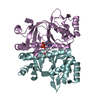
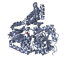
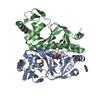
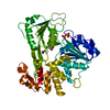
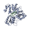
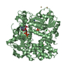
| Title | Structure of S. cerevisiae Hrr25:Mam1 complex, form 1 | ||||||
 Components Components |
| ||||||
 Keywords Keywords | TRANSFERASE / casein kinase / monopolin | ||||||
| Function / homology |  Function and homology information Function and homology informationmeiosis I sister chromatid cohesion / regulation of vesicle fusion with Golgi apparatus / regulation of protein localization by the Cvt pathway / monopolin complex / positive regulation of clathrin-dependent endocytosis / spindle attachment to meiosis I kinetochore / meiotic chromosome segregation / regulation of ER to Golgi vesicle-mediated transport / COPII-mediated vesicle transport / tRNA wobble uridine modification ...meiosis I sister chromatid cohesion / regulation of vesicle fusion with Golgi apparatus / regulation of protein localization by the Cvt pathway / monopolin complex / positive regulation of clathrin-dependent endocytosis / spindle attachment to meiosis I kinetochore / meiotic chromosome segregation / regulation of ER to Golgi vesicle-mediated transport / COPII-mediated vesicle transport / tRNA wobble uridine modification / homologous chromosome segregation / cellular bud tip / regulation of autophagosome assembly / cellular bud neck / pexophagy / spindle pole body / preribosome, small subunit precursor / chromosome, centromeric region / regulation of DNA repair / ribosomal large subunit biogenesis / P-body / kinetochore / endocytosis / regulation of protein localization / ribosomal small subunit biogenesis / protein tyrosine kinase activity / protein kinase activity / non-specific serine/threonine protein kinase / protein serine kinase activity / DNA repair / protein serine/threonine kinase activity / nucleolus / Golgi apparatus / signal transduction / nucleoplasm / ATP binding / identical protein binding / nucleus / plasma membrane / cytoplasm / cytosol Similarity search - Function | ||||||
| Biological species |  | ||||||
| Method |  X-RAY DIFFRACTION / SYNCHROTRON / MOLECULAR REPLACEMENT / Resolution: 1.841 Å X-RAY DIFFRACTION / SYNCHROTRON / MOLECULAR REPLACEMENT / Resolution: 1.841 Å | ||||||
 Authors Authors | Ye, Q. / Corbett, K.D. | ||||||
| Funding support |  United States, 1items United States, 1items
| ||||||
 Citation Citation | Journal: Embo J. / Year: 2016 Title: Structure of the Saccharomyces cerevisiae Hrr25:Mam1 monopolin subcomplex reveals a novel kinase regulator. Authors: Ye, Q. / Ur, S.N. / Su, T.Y. / Corbett, K.D. | ||||||
| History |
|
- Structure visualization
| Structure viewer | Molecule: MolmilJmol/JSmol |
|---|
- Downloads & links
-Download
| PDBx/mmCIF format | 5cyz.cif.gz | 222.3 KB | Display | PDBx/mmCIF format |
|---|---|---|---|---|
| PDB format | pdb5cyz.ent.gz | 176.5 KB | Display | PDB format |
| PDBx/mmJSON format | 5cyz.json.gz | Tree view | PDBx/mmJSON format | |
| Others |  Other downloads Other downloads |
-Validation report
| Arichive directory | https://data.pdbj.org/pub/pdb/validation_reports/cy/5cyzftp://data.pdbj.org/pub/pdb/validation_reports/cy/5cyz | HTTPS FTP |
|---|
-Related structure data
| Related structure data | 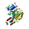 4xh0C 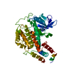 4xhgC 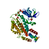 4xhhC 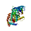 4xhlSC 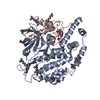 5czoC S: Starting model for refinement C: citing same article ( |
|---|---|
| Similar structure data |


-Links
 PDBj
PDBj
- Assembly
| Deposited unit | 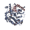
| ||||||||
|---|---|---|---|---|---|---|---|---|---|
| 1 |
| ||||||||
| Unit cell |
|
-Components
| #1: Protein | Mass: 46438.375 Da / Num. of mol.: 1 / Mutation: K38R, N-terminal Ala from tag Source method: isolated from a genetically manipulated source Details: surface LYS residues methylated (MLY) Source: (gene. exp.) Strain: ATCC 204508 / S288c / Gene: HRR25, YPL204W / Production host:  References: UniProt: P29295, non-specific serine/threonine protein kinase |
|---|---|
| #2: Protein | Mass: 12903.680 Da / Num. of mol.: 1 Source method: isolated from a genetically manipulated source Details: surface LYS residues methylated (MLY) Source: (gene. exp.) Strain: ATCC 204508 / S288c / Gene: MAM1, YER106W / Production host: |
| #3: Chemical | ChemComp-ZN /   Mass: 65.409 Da / Num. of mol.: 1 / Source method: obtained synthetically / Formula: Zn Mass: 65.409 Da / Num. of mol.: 1 / Source method: obtained synthetically / Formula: Zn |
| #4: Water | ChemComp-HOH /  Mass: 18.015 Da / Num. of mol.: 353 / Source method: isolated from a natural source / Formula: H2O Mass: 18.015 Da / Num. of mol.: 353 / Source method: isolated from a natural source / Formula: H2O |
-Experimental details
-Experiment
| Experiment | Method: X-RAY DIFFRACTION |
|---|
- Sample preparation
| Crystal | Density Matthews: 2.45 Å3/Da / Density % sol: 49.83 % |
|---|---|
| Crystal grow | Temperature: 293 K / Method: vapor diffusion, hanging drop / pH: 8.9 Details: 0.1M CHES pH8.9, 5% Tacsimate, 25 mM YCl3, 19% PEG3350 |
-Data collection
| Diffraction | Mean temperature: 100 K |
|---|---|
| Diffraction source | Source: SYNCHROTRON / Site: APS / Beamline: 24-ID-C / Wavelength: 0.979 Å |
| Detector | Type: PSI PILATUS 6M / Detector: PIXEL / Date: Jul 2, 2015 |
| Radiation | Protocol: SINGLE WAVELENGTH / Monochromatic (M) / Laue (L): M / Scattering type: x-ray |
| Radiation wavelength | Wavelength: 0.979 Å / Relative weight: 1 |
| Reflection | Resolution: 1.84→48.2 Å / Num. obs: 48597 / % possible obs: 99 % / Redundancy: 6.1 % / Rsym value: 0.152 / Net I/σ(I): 17 |
| Reflection shell | Resolution: 1.85→1.88 Å / Redundancy: 4.6 % / Mean I/σ(I) obs: 1.4 / % possible all: 98 |
- Processing
| Software |
| |||||||||||||||||||||||||||||||||||||||||||||||||||||||||||||||||||||||||||||||||||||||||||||||||||||||||||||||||||||||||||||||||||||
|---|---|---|---|---|---|---|---|---|---|---|---|---|---|---|---|---|---|---|---|---|---|---|---|---|---|---|---|---|---|---|---|---|---|---|---|---|---|---|---|---|---|---|---|---|---|---|---|---|---|---|---|---|---|---|---|---|---|---|---|---|---|---|---|---|---|---|---|---|---|---|---|---|---|---|---|---|---|---|---|---|---|---|---|---|---|---|---|---|---|---|---|---|---|---|---|---|---|---|---|---|---|---|---|---|---|---|---|---|---|---|---|---|---|---|---|---|---|---|---|---|---|---|---|---|---|---|---|---|---|---|---|---|---|---|
| Refinement | Method to determine structure: MOLECULAR REPLACEMENT Starting model: 4XHL Resolution: 1.841→48.2 Å / SU ML: 0.21 / Cross valid method: FREE R-VALUE / σ(F): 1.37 / Phase error: 19.96 / Stereochemistry target values: ML
| |||||||||||||||||||||||||||||||||||||||||||||||||||||||||||||||||||||||||||||||||||||||||||||||||||||||||||||||||||||||||||||||||||||
| Solvent computation | Shrinkage radii: 0.9 Å / VDW probe radii: 1.11 Å / Solvent model: FLAT BULK SOLVENT MODEL | |||||||||||||||||||||||||||||||||||||||||||||||||||||||||||||||||||||||||||||||||||||||||||||||||||||||||||||||||||||||||||||||||||||
| Refinement step | Cycle: LAST / Resolution: 1.841→48.2 Å
| |||||||||||||||||||||||||||||||||||||||||||||||||||||||||||||||||||||||||||||||||||||||||||||||||||||||||||||||||||||||||||||||||||||
| Refine LS restraints |
| |||||||||||||||||||||||||||||||||||||||||||||||||||||||||||||||||||||||||||||||||||||||||||||||||||||||||||||||||||||||||||||||||||||
| LS refinement shell |
| |||||||||||||||||||||||||||||||||||||||||||||||||||||||||||||||||||||||||||||||||||||||||||||||||||||||||||||||||||||||||||||||||||||
| Refinement TLS params. | Method: refined / Refine-ID: X-RAY DIFFRACTION
| |||||||||||||||||||||||||||||||||||||||||||||||||||||||||||||||||||||||||||||||||||||||||||||||||||||||||||||||||||||||||||||||||||||
| Refinement TLS group |
|
