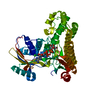
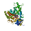
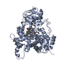
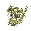
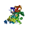
- PDB-4xhg: Structure of C. glabrata Hrr25 bound to ADP (formate condition) -
+
Open data
ID or keywords:
Loading...
-
Basic information
Entry
Database: PDB / ID: 4xhg
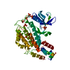
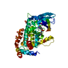
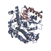
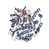
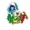
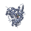
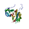
Title
Structure of C. glabrata Hrr25 bound to ADP (formate condition)
Components
Similar to uniprot|P29295 Saccharomyces cerevisiae YPL204w HRR25
Keywords
TRANSFERASE / casein kinase / monopolin
Function / homology
Function and homology information
regulation of vesicle fusion with Golgi apparatus / regulation of protein localization by the Cvt pathway / monopolin complex / positive regulation of clathrin-dependent endocytosis / spindle attachment to meiosis I kinetochore / regulation of ER to Golgi vesicle-mediated transport / tRNA wobble uridine modification / cellular bud tip / cellular bud neck / regulation of autophagosome assembly ...regulation of vesicle fusion with Golgi apparatus / regulation of protein localization by the Cvt pathway / monopolin complex / positive regulation of clathrin-dependent endocytosis / spindle attachment to meiosis I kinetochore / regulation of ER to Golgi vesicle-mediated transport / tRNA wobble uridine modification / cellular bud tip / cellular bud neck / regulation of autophagosome assembly / pexophagy / spindle pole body / preribosome, small subunit precursor / ribosomal large subunit biogenesis / P-body / ribosomal small subunit biogenesis / protein tyrosine kinase activity / non-specific serine/threonine protein kinase / DNA repair / protein serine/threonine kinase activity / Golgi apparatus / ATP binding / identical protein binding / nucleus / plasma membrane Similarity search - Function
: / Serine/threonine-protein kinase, active site / Serine/Threonine protein kinases active-site signature. / Protein kinase domain / Serine/Threonine protein kinases, catalytic domain / Protein kinase, ATP binding site / Protein kinases ATP-binding region signature. / Protein kinase domain profile. / Protein kinase domain / Protein kinase-like domain superfamily Similarity search - Domain/homology
In the structure databanks used in Yorodumi, some data are registered as the other names, "COVID-19 virus" and "2019-nCoV". Here are the details of the virus and the list of structure data.
Jan 31, 2019. EMDB accession codes are about to change! (news from PDBe EMDB page)
EMDB accession codes are about to change! (news from PDBe EMDB page)
The allocation of 4 digits for EMDB accession codes will soon come to an end. Whilst these codes will remain in use, new EMDB accession codes will include an additional digit and will expand incrementally as the available range of codes is exhausted. The current 4-digit format prefixed with “EMD-” (i.e. EMD-XXXX) will advance to a 5-digit format (i.e. EMD-XXXXX), and so on. It is currently estimated that the 4-digit codes will be depleted around Spring 2019, at which point the 5-digit format will come into force.
The EM Navigator/Yorodumi systems omit the EMD- prefix.
Related info.:Q: What is EMD? / ID/Accession-code notation in Yorodumi/EM Navigator
Yorodumi is a browser for structure data from EMDB, PDB, SASBDB, etc.
This page is also the successor to EM Navigator detail page, and also detail information page/front-end page for Omokage search.
The word "yorodu" (or yorozu) is an old Japanese word meaning "ten thousand". "mi" (miru) is to see.
Related info.:EMDB / PDB / SASBDB / Comparison of 3 databanks / Yorodumi Search / Aug 31, 2016. New EM Navigator & Yorodumi / Yorodumi Papers / Jmol/JSmol / Function and homology information / Changes in new EM Navigator and Yorodumi
 Movie
Movie Controller


 Open data
 Basic information
 Components
 Keywords
 Function and homology information
 Candida glabrata (fungus)
 X-RAY DIFFRACTION /
  Authors
 United States, 1items
  Citation
 Structure visualization
 Downloads & links
 Other downloads
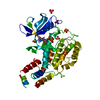


 PDBj
PDBj Assembly
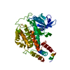


 Mass: 427.201 Da / Num. of mol.: 1 / Source method: obtained synthetically / Formula: C10H15N5O10P2 / Comment: ADP, energy-carrying molecule*YM
 Mass: 46.025 Da / Num. of mol.: 1 / Source method: obtained synthetically / Formula: CH2O2
 Mass: 18.015 Da / Num. of mol.: 285 / Source method: isolated from a natural source / Formula: H2O
 Sample preparation
 Processing