 Movie
Movie Controller

[English] 日本語
 Yorodumi
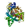
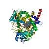
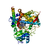
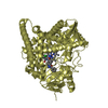
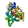
- PDB-2wx2: X-RAY STRUCTURE OF CYP51 FROM THE HUMAN PATHOGEN TRYPANOSOMA CRUZ... -
+ Open data

- Basic information
| Entry | Database: PDB / ID: 2wx2 | ||||||
|---|---|---|---|---|---|---|---|
| Title | X-RAY STRUCTURE OF CYP51 FROM THE HUMAN PATHOGEN TRYPANOSOMA CRUZI IN COMPLEX WITH FLUCONAZOLE | ||||||
 Components Components | LANOSTEROL 14-ALPHA-DEMETHYLASE | ||||||
 Keywords Keywords | OXIDOREDUCTASE / P450 / HEME / IRON / METAL-BINDING / METHYLTRANSFERASE / ERGOSTEROL BIOSYNTHESIS | ||||||
| Function / homology |  Function and homology information Function and homology informationsterol biosynthetic process / sterol 14alpha-demethylase / sterol 14-demethylase activity / iron ion binding / heme binding / membrane Similarity search - Function | ||||||
| Biological species |  | ||||||
| Method |  X-RAY DIFFRACTION / SYNCHROTRON / MOLECULAR REPLACEMENT / Resolution: 2.27 Å X-RAY DIFFRACTION / SYNCHROTRON / MOLECULAR REPLACEMENT / Resolution: 2.27 Å | ||||||
 Authors Authors | Chen, C.-K. / Leung, S.S.F. / Guilbert, C. / Jacobson, M. / McKerrow, J.H. / Podust, L.M. | ||||||
 Citation Citation | Journal: Plos Negl Trop Dis / Year: 2010 Title: Structural Characterization of Cyp51 from Trypanosoma Cruzi and Trypanosoma Brucei Bound to the Antifungal Drugs Posaconazole and Fluconazole Authors: Chen, C.-K. / Leung, S.S.F. / Guilbert, C. / Jacobson, M. / Mckerrow, J.H. / Podust, L.M. | ||||||
| History |
|
- Structure visualization
| Structure viewer | Molecule: MolmilJmol/JSmol |
|---|
- Downloads & links
-Download
| PDBx/mmCIF format | 2wx2.cif.gz | 367 KB | Display | PDBx/mmCIF format |
|---|---|---|---|---|
| PDB format | pdb2wx2.ent.gz | 296.6 KB | Display | PDB format |
| PDBx/mmJSON format | 2wx2.json.gz | Tree view | PDBx/mmJSON format | |
| Others |  Other downloads Other downloads |
-Validation report
| Arichive directory | https://data.pdbj.org/pub/pdb/validation_reports/wx/2wx2ftp://data.pdbj.org/pub/pdb/validation_reports/wx/2wx2 | HTTPS FTP |
|---|
-Related structure data
| Related structure data | 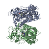 2wuzSC 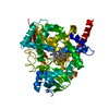 2wv2C 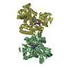 2x2nC C: citing same article ( S: Starting model for refinement |
|---|---|
| Similar structure data |


-Links
 PDBj
PDBj
- Assembly
| Deposited unit | 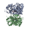
| ||||||||
|---|---|---|---|---|---|---|---|---|---|
| 1 | 
| ||||||||
| 2 | 
| ||||||||
| Unit cell |
| ||||||||
| Noncrystallographic symmetry (NCS) | NCS oper: (Code: given Matrix: (0.53951, -0.27526, 0.79571), Vector: |
-Components
| #1: Protein | Mass: 54120.766 Da / Num. of mol.: 2 / Fragment: RESIDUES 22-481 Source method: isolated from a genetically manipulated source Details: FIRST 20 RESIDUES UPSTREAM OF K21 WERE REPLACED WITH THE FRAGMENT MAKKKKK Source: (gene. exp.)  #2: Chemical |   Mass: 616.487 Da / Num. of mol.: 2 / Source method: obtained synthetically / Formula: C34H32FeN4O4 Mass: 616.487 Da / Num. of mol.: 2 / Source method: obtained synthetically / Formula: C34H32FeN4O4#3: Chemical | 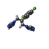  Mass: 306.271 Da / Num. of mol.: 2 / Source method: obtained synthetically / Formula: C13H12F2N6O / Comment: medication*YM Mass: 306.271 Da / Num. of mol.: 2 / Source method: obtained synthetically / Formula: C13H12F2N6O / Comment: medication*YM#4: Water | ChemComp-HOH / |  Mass: 18.015 Da / Num. of mol.: 429 / Source method: isolated from a natural source / Formula: H2O Mass: 18.015 Da / Num. of mol.: 429 / Source method: isolated from a natural source / Formula: H2ONonpolymer details | PROTOPORPHYRIN IX CONTAINING FE (HEM): THIOLATE BOND BETWEEN THE HEME IRON AND CYS 422 FLUCONAZOLE ...PROTOPORPH | Sequence details | THE FIRST 21 RESIDUES UPSTREAM OF K22 WERE REPLACED WITH THE FRAGMENT MAKKKKK AT THE N-TERMINUS. ...THE FIRST 21 RESIDUES UPSTREAM OF K22 WERE REPLACED WITH THE FRAGMENT MAKKKKK AT THE N-TERMINUS. HIS6-TAG WAS INTRODUCED | |
|---|
-Experimental details
-Experiment
| Experiment | Method: X-RAY DIFFRACTION / Number of used crystals: 1 |
|---|
- Sample preparation
| Crystal | Density Matthews: 2.3 Å3/Da / Density % sol: 47 % / Description: NONE |
|---|---|
| Crystal grow | pH: 5.5 / Details: 25% PEG 4000, 0.1 M BIS-TRIS, PH 5.5 |
-Data collection
| Diffraction | Mean temperature: 110 K |
|---|---|
| Diffraction source | Source: SYNCHROTRON / Site: ALS  / Beamline: 8.3.1 / Wavelength: 1.11587 / Beamline: 8.3.1 / Wavelength: 1.11587 |
| Detector | Type: ADSC CCD / Detector: CCD / Date: Oct 23, 2009 / Details: MIRRORS |
| Radiation | Monochromator: SI (111) DOUBLE CRYSTAL / Protocol: SINGLE WAVELENGTH / Monochromatic (M) / Laue (L): M / Scattering type: x-ray |
| Radiation wavelength | Wavelength: 1.11587 Å / Relative weight: 1 |
| Reflection | Resolution: 2.27→24.3 Å / Num. obs: 40051 / % possible obs: 100 % / Observed criterion σ(I): 0 / Redundancy: 3.6 % / Biso Wilson estimate: 31.8 Å2 / Rmerge(I) obs: 0.09 / Net I/σ(I): 8.5 |
| Reflection shell | Resolution: 2.27→2.39 Å / Redundancy: 2.4 % / Rmerge(I) obs: 0.42 / Mean I/σ(I) obs: 2.4 / % possible all: 55.7 |
- Processing
| Software |
| ||||||||||||||||||||||||||||||||||||||||||||||||||||||||||||||||||||||||||||||||||||||||||||||||||||||||||||||||||||||||||||||||||||||||||||||||||||||||||||||||||||||||||||||||||||||
|---|---|---|---|---|---|---|---|---|---|---|---|---|---|---|---|---|---|---|---|---|---|---|---|---|---|---|---|---|---|---|---|---|---|---|---|---|---|---|---|---|---|---|---|---|---|---|---|---|---|---|---|---|---|---|---|---|---|---|---|---|---|---|---|---|---|---|---|---|---|---|---|---|---|---|---|---|---|---|---|---|---|---|---|---|---|---|---|---|---|---|---|---|---|---|---|---|---|---|---|---|---|---|---|---|---|---|---|---|---|---|---|---|---|---|---|---|---|---|---|---|---|---|---|---|---|---|---|---|---|---|---|---|---|---|---|---|---|---|---|---|---|---|---|---|---|---|---|---|---|---|---|---|---|---|---|---|---|---|---|---|---|---|---|---|---|---|---|---|---|---|---|---|---|---|---|---|---|---|---|---|---|---|---|
| Refinement | Method to determine structure: MOLECULAR REPLACEMENT Starting model: PDB ENTRY 2WUZ Resolution: 2.27→69.5 Å / Cor.coef. Fo:Fc: 0.936 / Cor.coef. Fo:Fc free: 0.872 / SU B: 18.013 / SU ML: 0.201 / Cross valid method: THROUGHOUT / ESU R Free: 0.291 / Stereochemistry target values: MAXIMUM LIKELIHOOD Details: HYDROGENS HAVE BEEN ADDED IN THE RIDING POSITIONS. DISORDERED REGIONS WERE OMITTED FROM THE STRUCTURE
| ||||||||||||||||||||||||||||||||||||||||||||||||||||||||||||||||||||||||||||||||||||||||||||||||||||||||||||||||||||||||||||||||||||||||||||||||||||||||||||||||||||||||||||||||||||||
| Solvent computation | Ion probe radii: 0.8 Å / Shrinkage radii: 0.8 Å / VDW probe radii: 1.4 Å / Solvent model: MASK | ||||||||||||||||||||||||||||||||||||||||||||||||||||||||||||||||||||||||||||||||||||||||||||||||||||||||||||||||||||||||||||||||||||||||||||||||||||||||||||||||||||||||||||||||||||||
| Displacement parameters | Biso mean: 23.751 Å2
| ||||||||||||||||||||||||||||||||||||||||||||||||||||||||||||||||||||||||||||||||||||||||||||||||||||||||||||||||||||||||||||||||||||||||||||||||||||||||||||||||||||||||||||||||||||||
| Refinement step | Cycle: LAST / Resolution: 2.27→69.5 Å
| ||||||||||||||||||||||||||||||||||||||||||||||||||||||||||||||||||||||||||||||||||||||||||||||||||||||||||||||||||||||||||||||||||||||||||||||||||||||||||||||||||||||||||||||||||||||
| Refine LS restraints |
|