 Movie
Movie Controller

[English] 日本語
 Yorodumi
- PDB-4g7g: Sterol 14-alpha demethylase (CYP51) from Trypanosoma brucei in co... -
+ Open data

- Basic information
| Entry | Database: PDB / ID: 4g7g | ||||||
|---|---|---|---|---|---|---|---|
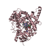
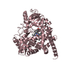
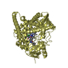
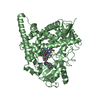
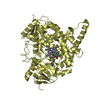
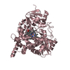
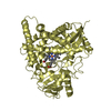
| Title | Sterol 14-alpha demethylase (CYP51) from Trypanosoma brucei in complex with the VNI derivative (R)-N-(1-(3,4'-difluorobiphenyl-4-yl)-2-(1H-imidazol-1-yl)ethyl)-4-(5-phenyl-1,3,4-oxadiazol-2-yl)benzamide [VNI/VNF (VFV)] | ||||||
 Components Components | sterol 14-alpha-demethylase | ||||||
 Keywords Keywords | OXIDOREDUCTASE/OXIDOREDUCTASE INHIBITOR / cytochrome P450 fold / heme / monooxygenase / sterol biosynthesis / eukaryotic membrane biogenesis / cytochrome P450 reductase / endoplasmic reticulum membrane / OXIDOREDUCTASE-OXIDOREDUCTASE INHIBITOR complex | ||||||
| Function / homology |  Function and homology information Function and homology informationmembrane biogenesis / sterol 14alpha-demethylase / sterol 14-demethylase activity / nuclear envelope / oxidoreductase activity / iron ion binding / heme binding / endoplasmic reticulum Similarity search - Function | ||||||
| Biological species |  | ||||||
| Method |  X-RAY DIFFRACTION / SYNCHROTRON / MOLECULAR REPLACEMENT / Resolution: 2.05 Å X-RAY DIFFRACTION / SYNCHROTRON / MOLECULAR REPLACEMENT / Resolution: 2.05 Å | ||||||
 Authors Authors | Hargrove, T.Y. / Wawrzak, Z. / Waterman, M.R. / Lepesheva, G.I. | ||||||
 Citation Citation | Journal: J Infect Dis / Year: 2015 Title: VFV as a New Effective CYP51 Structure-Derived Drug Candidate for Chagas Disease and Visceral Leishmaniasis. Authors: Lepesheva, G.I. / Hargrove, T.Y. / Rachakonda, G. / Wawrzak, Z. / Pomel, S. / Cojean, S. / Nde, P.N. / Nes, W.D. / Locuson, C.W. / Calcutt, M.W. / Waterman, M.R. / Daniels, J.S. / Loiseau, P.M. / Villalta, F. | ||||||
| History |
|
- Structure visualization
| Structure viewer | Molecule: MolmilJmol/JSmol |
|---|
- Downloads & links
-Download
| PDBx/mmCIF format | 4g7g.cif.gz | 747.8 KB | Display | PDBx/mmCIF format |
|---|---|---|---|---|
| PDB format | pdb4g7g.ent.gz | 620.2 KB | Display | PDB format |
| PDBx/mmJSON format | 4g7g.json.gz | Tree view | PDBx/mmJSON format | |
| Others |  Other downloads Other downloads |
-Validation report
| Arichive directory | https://data.pdbj.org/pub/pdb/validation_reports/g7/4g7gftp://data.pdbj.org/pub/pdb/validation_reports/g7/4g7g | HTTPS FTP |
|---|
-Related structure data
| Related structure data |  4g3jC  3g1qS C: citing same article ( S: Starting model for refinement |
|---|---|
| Similar structure data |


-Links
 PDBj
PDBj
- Assembly
| Deposited unit | 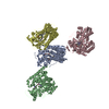
| ||||||||
|---|---|---|---|---|---|---|---|---|---|
| 1 | 
| ||||||||
| 2 | 
| ||||||||
| 3 | 
| ||||||||
| 4 | 
| ||||||||
| Unit cell |
|
-Components
| #1: Protein | Mass: 50759.902 Da / Num. of mol.: 4 / Fragment: UNP residues 29-476 / Mutation: P29G, T30K, D31L Source method: isolated from a genetically manipulated source Source: (gene. exp.)  #2: Chemical | ChemComp-HEM /   Mass: 616.487 Da / Num. of mol.: 4 / Source method: obtained synthetically / Formula: C34H32FeN4O4 Mass: 616.487 Da / Num. of mol.: 4 / Source method: obtained synthetically / Formula: C34H32FeN4O4#3: Chemical | ChemComp-VFV /   Mass: 547.554 Da / Num. of mol.: 4 / Source method: obtained synthetically / Formula: C32H23F2N5O2 Mass: 547.554 Da / Num. of mol.: 4 / Source method: obtained synthetically / Formula: C32H23F2N5O2#4: Water | ChemComp-HOH / |  Mass: 18.015 Da / Num. of mol.: 504 / Source method: isolated from a natural source / Formula: H2O Mass: 18.015 Da / Num. of mol.: 504 / Source method: isolated from a natural source / Formula: H2O |
|---|
-Experimental details
-Experiment
| Experiment | Method: X-RAY DIFFRACTION / Number of used crystals: 1 |
|---|
- Sample preparation
| Crystal | Density Matthews: 2.49 Å3/Da / Density % sol: 50.63 % |
|---|---|
| Crystal grow | Temperature: 294 K / Method: vapor diffusion, hanging drop / pH: 7.3 Details: potassium phosphate, sodium chloride, glycerol, PEG3350, N-tetradecyl-beta-D-maltoside, pH 7.3, VAPOR DIFFUSION, HANGING DROP, temperature 294K |
-Data collection
| Diffraction | Mean temperature: 100 K |
|---|---|
| Diffraction source | Source: SYNCHROTRON / Site: APS  / Beamline: 21-ID-F / Wavelength: 0.97872 Å / Beamline: 21-ID-F / Wavelength: 0.97872 Å |
| Detector | Type: MARMOSAIC 225 mm CCD / Detector: CCD / Date: Apr 3, 2012 / Details: Be Lenses/Diamond Laue Mono |
| Radiation | Monochromator: Diamond(111) / Protocol: SINGLE WAVELENGTH / Monochromatic (M) / Laue (L): M / Scattering type: x-ray |
| Radiation wavelength | Wavelength: 0.97872 Å / Relative weight: 1 |
| Reflection | Resolution: 2.05→29.817 Å / Num. all: 123297 / Num. obs: 120831 / % possible obs: 98 % / Observed criterion σ(F): 2.6 / Observed criterion σ(I): 2.6 / Redundancy: 4.6 % / Biso Wilson estimate: 37.85 Å2 / Rmerge(I) obs: 0.046 / Rsym value: 0.046 / Net I/σ(I): 31 |
| Reflection shell | Resolution: 2.05→2.09 Å / Redundancy: 4.5 % / Rmerge(I) obs: 0.58 / Mean I/σ(I) obs: 2.6 / Num. unique all: 5970 / % possible all: 97 |
- Processing
| Software |
| ||||||||||||||||||||||||||||||||||||||||||||||||||||||||||||
|---|---|---|---|---|---|---|---|---|---|---|---|---|---|---|---|---|---|---|---|---|---|---|---|---|---|---|---|---|---|---|---|---|---|---|---|---|---|---|---|---|---|---|---|---|---|---|---|---|---|---|---|---|---|---|---|---|---|---|---|---|---|
| Refinement | Method to determine structure: MOLECULAR REPLACEMENT Starting model: PDB ENTRY 3G1Q Resolution: 2.05→29.817 Å / Cor.coef. Fo:Fc: 0.972 / Cor.coef. Fo:Fc free: 0.95 / SU B: 11.406 / SU ML: 0.137 / Cross valid method: THROUGHOUT / ESU R Free: 0.172 / Stereochemistry target values: MAXIMUM LIKELIHOOD Details: HYDROGENS HAVE BEEN USED IF PRESENT IN THE INPUT U VALUES : REFINED INDIVIDUALLY
| ||||||||||||||||||||||||||||||||||||||||||||||||||||||||||||
| Solvent computation | Ion probe radii: 0.8 Å / Shrinkage radii: 0.8 Å / VDW probe radii: 1.2 Å / Solvent model: MASK | ||||||||||||||||||||||||||||||||||||||||||||||||||||||||||||
| Displacement parameters | Biso mean: 44.796 Å2
| ||||||||||||||||||||||||||||||||||||||||||||||||||||||||||||
| Refinement step | Cycle: LAST / Resolution: 2.05→29.817 Å
| ||||||||||||||||||||||||||||||||||||||||||||||||||||||||||||
| Refine LS restraints |
| ||||||||||||||||||||||||||||||||||||||||||||||||||||||||||||
| LS refinement shell | Resolution: 2.05→2.102 Å / Total num. of bins used: 20
|
