 Movie
Movie Controller

[English] 日本語
 Yorodumi
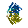
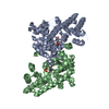
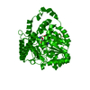
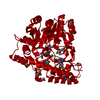
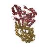
- PDB-1m4n: CRYSTAL STRUCTURE OF APPLE ACC SYNTHASE IN COMPLEX WITH [2-(AMINO... -
+ Open data

- Basic information
| Entry | Database: PDB / ID: 1m4n | ||||||
|---|---|---|---|---|---|---|---|
| Title | CRYSTAL STRUCTURE OF APPLE ACC SYNTHASE IN COMPLEX WITH [2-(AMINO-OXY)ETHYL](5'-DEOXYADENOSIN-5'-YL)(METHYL)SULFONIUM | ||||||
 Components Components | 1-aminocyclopropane-1-carboxylate synthase | ||||||
 Keywords Keywords | LYASE / Fruit ripening / Ethylene biosynthesis / Pyridoxal phosphate | ||||||
| Function / homology |  Function and homology information Function and homology informationOxidoreductases; Acting on the CH-NH2 group of donors / 1-aminocyclopropane-1-carboxylate synthase / 1-aminocyclopropane-1-carboxylate synthase activity / fruit ripening / ethylene biosynthetic process / transaminase activity / pyridoxal phosphate binding / oxidoreductase activity Similarity search - Function | ||||||
| Biological species |  | ||||||
| Method |  X-RAY DIFFRACTION / MOLECULAR REPLACEMENT / Resolution: 2.01 Å X-RAY DIFFRACTION / MOLECULAR REPLACEMENT / Resolution: 2.01 Å | ||||||
 Authors Authors | Capitani, G. / Eliot, A.C. / Gut, H. / Khomutov, R.M. / Kirsch, J.F. / Grutter, M.G. | ||||||
 Citation Citation | Journal: BIOCHEM.BIOPHYS.ACTA PROTEINS & PROTEOMICS / Year: 2003 Title: Structure of 1-aminocyclopropane-1-carboxylate synthase in complex with an amino-oxy analogue of the substrate: implications for substrate binding. Authors: Capitani, G. / Eliot, A.C. / Gut, H. / Khomutov, R.M. / Kirsch, J.F. / Grutter, M.G. | ||||||
| History |
|
- Structure visualization
| Structure viewer | Molecule: MolmilJmol/JSmol |
|---|
- Downloads & links
-Download
| PDBx/mmCIF format | 1m4n.cif.gz | 105.7 KB | Display | PDBx/mmCIF format |
|---|---|---|---|---|
| PDB format | pdb1m4n.ent.gz | 78.7 KB | Display | PDB format |
| PDBx/mmJSON format | 1m4n.json.gz | Tree view | PDBx/mmJSON format | |
| Others |  Other downloads Other downloads |
-Validation report
| Arichive directory | https://data.pdbj.org/pub/pdb/validation_reports/m4/1m4nftp://data.pdbj.org/pub/pdb/validation_reports/m4/1m4n | HTTPS FTP |
|---|
-Related structure data
| Related structure data |  1b8gS S: Starting model for refinement |
|---|---|
| Similar structure data |


-Links
 PDBj
PDBj- Assembly
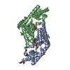
| Deposited unit | 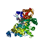
| ||||||||||||
|---|---|---|---|---|---|---|---|---|---|---|---|---|---|
| 1 | 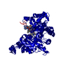
| ||||||||||||
| Unit cell |
| ||||||||||||
| Components on special symmetry positions |
| ||||||||||||
| Details | The biological assembly is a dimer |
-Components
| #1: Protein | Mass: 49091.797 Da / Num. of mol.: 1 / Fragment: RESIDUES 1-435 Source method: isolated from a genetically manipulated source Source: (gene. exp.)  References: UniProt: P37821, 1-aminocyclopropane-1-carboxylate synthase |
|---|---|
| #2: Chemical | ChemComp-PLP /   Mass: 247.142 Da / Num. of mol.: 1 / Source method: obtained synthetically / Formula: C8H10NO6P Mass: 247.142 Da / Num. of mol.: 1 / Source method: obtained synthetically / Formula: C8H10NO6P |
| #3: Chemical | ChemComp-AAD / (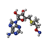  Mass: 357.409 Da / Num. of mol.: 1 / Source method: obtained synthetically / Formula: C13H21N6O4S Mass: 357.409 Da / Num. of mol.: 1 / Source method: obtained synthetically / Formula: C13H21N6O4S |
| #4: Chemical | ChemComp-MES /   Mass: 195.237 Da / Num. of mol.: 1 / Source method: obtained synthetically / Formula: C6H13NO4S / Comment: pH buffer*YM Mass: 195.237 Da / Num. of mol.: 1 / Source method: obtained synthetically / Formula: C6H13NO4S / Comment: pH buffer*YM |
| #5: Water | ChemComp-HOH /  Mass: 18.015 Da / Num. of mol.: 288 / Source method: isolated from a natural source / Formula: H2O Mass: 18.015 Da / Num. of mol.: 288 / Source method: isolated from a natural source / Formula: H2O |
-Experimental details
-Experiment
| Experiment | Method: X-RAY DIFFRACTION / Number of used crystals: 1 |
|---|
- Sample preparation
| Crystal | Density Matthews: 2.1 Å3/Da / Density % sol: 41.53 % | ||||||||||||||||||||||||||||||||||||||||||||||||
|---|---|---|---|---|---|---|---|---|---|---|---|---|---|---|---|---|---|---|---|---|---|---|---|---|---|---|---|---|---|---|---|---|---|---|---|---|---|---|---|---|---|---|---|---|---|---|---|---|---|
| Crystal grow | Temperature: 293 K / Method: vapor diffusion, sitting drop / pH: 6.5 Details: MPD, MES, pH 6.5, VAPOR DIFFUSION, SITTING DROP, temperature 293K | ||||||||||||||||||||||||||||||||||||||||||||||||
| Crystal grow | *PLUS pH: 7.9 | ||||||||||||||||||||||||||||||||||||||||||||||||
| Components of the solutions | *PLUS
|
-Data collection
| Diffraction | Mean temperature: 100 K |
|---|---|
| Diffraction source | Source: ROTATING ANODE / Type: ENRAF-NONIUS / Wavelength: 1.5418 Å |
| Detector | Type: MARRESEARCH / Detector: IMAGE PLATE / Date: Apr 30, 2000 / Details: double focussing mirrors (Prophysics) |
| Radiation | Monochromator: MIRRORS (PROPHYSICS) + Ni FILTER / Protocol: SINGLE WAVELENGTH / Monochromatic (M) / Laue (L): M / Scattering type: x-ray |
| Radiation wavelength | Wavelength: 1.5418 Å / Relative weight: 1 |
| Reflection | Resolution: 2.01→23.77 Å / Num. obs: 26106 / % possible obs: 96.2 % / Observed criterion σ(I): -3 / Redundancy: 2.8 % / Biso Wilson estimate: 23.6 Å2 / Rmerge(I) obs: 0.077 / Net I/σ(I): 10.6 |
| Reflection shell | Resolution: 2.01→2.06 Å / Rmerge(I) obs: 0.29 / Mean I/σ(I) obs: 2 / Num. unique all: 1185 / % possible all: 66.5 |
| Reflection | *PLUS Lowest resolution: 24 Å |
| Reflection shell | *PLUS % possible obs: 66.5 % / Rmerge(I) obs: 0.29 |
- Processing
| Software |
| ||||||||||||||||||||
|---|---|---|---|---|---|---|---|---|---|---|---|---|---|---|---|---|---|---|---|---|---|
| Refinement | Method to determine structure: MOLECULAR REPLACEMENT Starting model: PDB ENTRY 1B8G Resolution: 2.01→23.77 Å / Isotropic thermal model: ISOTROPIC / Cross valid method: THROUGHOUT / σ(F): 0 / Stereochemistry target values: Engh & Huber / Details: Used bulk solvent correction (CNS 1.0)
| ||||||||||||||||||||
| Displacement parameters | Biso mean: 24.8 Å2
| ||||||||||||||||||||
| Refine analyze |
| ||||||||||||||||||||
| Refinement step | Cycle: LAST / Resolution: 2.01→23.77 Å
| ||||||||||||||||||||
| Refine LS restraints |
| ||||||||||||||||||||
| LS refinement shell | Resolution: 2.01→2.08 Å
| ||||||||||||||||||||
| Refinement | *PLUS Lowest resolution: 24 Å | ||||||||||||||||||||
| Solvent computation | *PLUS | ||||||||||||||||||||
| Displacement parameters | *PLUS | ||||||||||||||||||||
| Refine LS restraints | *PLUS
| ||||||||||||||||||||
| LS refinement shell | *PLUS Lowest resolution: 2.06 Å |
