 Movie
Movie Controller


+ Open data

- Basic information
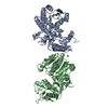
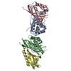
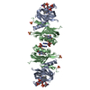
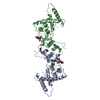
| Entry | Database: PDB / ID: 1bmo | |||||||||
|---|---|---|---|---|---|---|---|---|---|---|
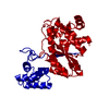
| Title | BM-40, FS/EC DOMAIN PAIR | |||||||||
 Components Components | BM-40 | |||||||||
 Keywords Keywords | EXTRACELLULAR MODULE /  GLYCOPROTEIN / ANTI-ADHESIVE PROTEIN GLYCOPROTEIN / ANTI-ADHESIVE PROTEIN | |||||||||
| Function / homology |  Function and homology information Function and homology informationScavenging by Class H Receptors / platelet alpha granule membrane / platelet alpha granule / anatomical structure development / extracellular matrix binding / regulation of cell morphogenesis / negative regulation of endothelial cell proliferation / basement membrane / ECM proteoglycans / Nuclear signaling by ERBB4 ...Scavenging by Class H Receptors / platelet alpha granule membrane / platelet alpha granule / anatomical structure development / extracellular matrix binding / regulation of cell morphogenesis / negative regulation of endothelial cell proliferation / basement membrane / ECM proteoglycans / Nuclear signaling by ERBB4 / endocytic vesicle lumen / collagen binding / negative regulation of angiogenesis / positive regulation of endothelial cell migration / platelet alpha granule lumen / nuclear matrix / Platelet degranulation / collagen-containing extracellular matrix / intracellular membrane-bounded organelle / calcium ion binding / cell surface / extracellular space / extracellular region / plasma membrane / cytoplasmSimilarity search - Function | |||||||||
| Biological species |  Homo sapiens (human) Homo sapiens (human) | |||||||||
| Method | X-RAY DIFFRACTION / MOLECULAR REPLACEMENT, 2-FOLD NCS AVERAGING / Resolution: 3.1 Å | |||||||||
 Authors Authors | Hohenester, E. / Maurer, P. / Timpl, R. | |||||||||
 Citation Citation | Journal: EMBO J. / Year: 1997 Title: Crystal structure of a pair of follistatin-like and EF-hand calcium-binding domains in BM-40. Authors: Hohenester, E. / Maurer, P. / Timpl, R. #1: Journal: Nat.Struct.Biol. / Year: 1996Title: Structure of a Novel Extracellular Ca(2+)-Binding Module in Bm-40 Authors: Hohenester, E. / Maurer, P. / Hohenadl, C. / Timpl, R. / Jansonius, J.N. / Engel, J. #2: Journal: J.Mol.Biol. / Year: 1995Title: The C-Terminal Portion of Bm-40 (Sparc/Osteonectin) is an Autonomously Folding and Crystallisable Domain that Binds Calcium and Collagen Iv Authors: Maurer, P. / Hohenadl, C. / Hohenester, E. / Gohring, W. / Timpl, R. / Engel, J. #3: Journal: Faseb J. / Year: 1994Title: The Biology of Sparc, a Protein that Modulates Cell-Matrix Interactions Authors: Lane, T.F. / Sage, E.H. | |||||||||
| History |
|
- Structure visualization
| Structure viewer | Molecule: MolmilJmol/JSmol |
|---|
- Downloads & links
-Download
| PDBx/mmCIF format | 1bmo.cif.gz | 105.8 KB | Display | PDBx/mmCIF format |
|---|---|---|---|---|
| PDB format | pdb1bmo.ent.gz | 83.3 KB | Display | PDB format |
| PDBx/mmJSON format | 1bmo.json.gz | Tree view | PDBx/mmJSON format | |
| Others |  Other downloads Other downloads |
-Validation report
| Arichive directory | https://data.pdbj.org/pub/pdb/validation_reports/bm/1bmoftp://data.pdbj.org/pub/pdb/validation_reports/bm/1bmo | HTTPS FTP |
|---|
-Related structure data
| Related structure data |  1sraS S: Starting model for refinement |
|---|---|
| Similar structure data |


-Links
 PDBj
PDBj


- Assembly
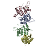
| Deposited unit | 
| |||||||||||||||||||||
|---|---|---|---|---|---|---|---|---|---|---|---|---|---|---|---|---|---|---|---|---|---|---|
| 1 |
| |||||||||||||||||||||
| Unit cell |
| |||||||||||||||||||||
| Noncrystallographic symmetry (NCS) | NCS domain:
NCS oper:
|
-Components
| #1: Protein | Mass: 27111.934 Da / Num. of mol.: 2 / Fragment: FS/EC DOMAIN PAIR, RESIDUES 53 - 286 Source method: isolated from a genetically manipulated source Source: (gene. exp.) Homo sapiens (human) / Cell line: 293 / Organ: KIDNEY / Cell line (production host): HEK293 / Production host: Homo sapiens (human) / References: UniProt: P09486#2: Polysaccharide | / Mass: 424.401 Da / Num. of mol.: 2Source method: isolated from a genetically manipulated source #3: Chemical | ChemComp-CA /   Mass: 40.078 Da / Num. of mol.: 4 / Source method: obtained synthetically / Formula: Ca Mass: 40.078 Da / Num. of mol.: 4 / Source method: obtained synthetically / Formula: Ca |
|---|
-Experimental details
-Experiment
| Experiment | Method: X-RAY DIFFRACTION / Number of used crystals: 2 |
|---|
- Sample preparation
| Crystal | Density Matthews: 4 Å3/Da / Density % sol: 65 % Description: THE R-FACTOR AFTER RIGID-BODY REFINEMENT OF THE TWO EC DOMAINS WAS 0.46 (8-3.1 A) | ||||||||||||||||||||||||||||||||||||||||||||||||||||||||||||
|---|---|---|---|---|---|---|---|---|---|---|---|---|---|---|---|---|---|---|---|---|---|---|---|---|---|---|---|---|---|---|---|---|---|---|---|---|---|---|---|---|---|---|---|---|---|---|---|---|---|---|---|---|---|---|---|---|---|---|---|---|---|
| Crystal grow | Method: vapor diffusion, hanging drop / pH: 7.5 Details: HANGING DROP VAPOUR DIFFUSION AT 20 DEG C: 10 MG/ML PROTEIN IN 10 MM TRIS PH 7.5 + 2 MM CACL2; 0.1 M HEPES PH 7.5, 0.15 M NA-ACETATE, 15% PEG4K, vapor diffusion - hanging drop | ||||||||||||||||||||||||||||||||||||||||||||||||||||||||||||
| Crystal grow | *PLUS Method: vapor diffusion, hanging drop | ||||||||||||||||||||||||||||||||||||||||||||||||||||||||||||
| Components of the solutions | *PLUS
|
-Data collection
| Diffraction | Mean temperature: 100 K |
|---|---|
| Diffraction source | Source: ROTATING ANODE / Type: ELLIOTT GX-21 / Wavelength: 1.5418 |
| Detector | Type: MARRESEARCH / Detector: IMAGE PLATE / Date: Oct 1, 1996 |
| Radiation | Monochromatic (M) / Laue (L): M / Scattering type: x-ray |
| Radiation wavelength | Wavelength: 1.5418 Å / Relative weight: 1 |
| Reflection | Resolution: 3.1→25 Å / Num. obs: 14328 / % possible obs: 90.7 % / Observed criterion σ(I): 0 / Redundancy: 3.3 % / Rmerge(I) obs: 0.086 / Net I/σ(I): 6.5 |
| Reflection shell | Resolution: 3.1→3.21 Å / Redundancy: 2.3 % / Rmerge(I) obs: 0.267 / Mean I/σ(I) obs: 2.7 / % possible all: 85.1 |
| Reflection | *PLUS Num. measured all: 47563 |
| Reflection shell | *PLUS % possible obs: 85.1 % |
- Processing
| Software |
| ||||||||||||||||||||||||||||||||||||||||||||||||||||||||||||||||||||||||||||||||
|---|---|---|---|---|---|---|---|---|---|---|---|---|---|---|---|---|---|---|---|---|---|---|---|---|---|---|---|---|---|---|---|---|---|---|---|---|---|---|---|---|---|---|---|---|---|---|---|---|---|---|---|---|---|---|---|---|---|---|---|---|---|---|---|---|---|---|---|---|---|---|---|---|---|---|---|---|---|---|---|---|---|
| Refinement | Method to determine structure: MOLECULAR REPLACEMENT, 2-FOLD NCS AVERAGING Starting model: BM-40 EC DOMAIN (PDB ENTRY 1SRA) Resolution: 3.1→8 Å / Data cutoff high absF: 100000 / Data cutoff low absF: 0.01 Isotropic thermal model: RESTRAINED INDIVIDUAL WIT NCS RESTRAINTS Cross valid method: FREE R-FACTOR / σ(F): 0 Details: A BULK SOLVENT CORRECTION WAS USED (R=0.25, K=0.33, B=50A**2)
| ||||||||||||||||||||||||||||||||||||||||||||||||||||||||||||||||||||||||||||||||
| Displacement parameters | Biso mean: 28 Å2 | ||||||||||||||||||||||||||||||||||||||||||||||||||||||||||||||||||||||||||||||||
| Refine analyze |
| ||||||||||||||||||||||||||||||||||||||||||||||||||||||||||||||||||||||||||||||||
| Refinement step | Cycle: LAST / Resolution: 3.1→8 Å
| ||||||||||||||||||||||||||||||||||||||||||||||||||||||||||||||||||||||||||||||||
| Refine LS restraints |
| ||||||||||||||||||||||||||||||||||||||||||||||||||||||||||||||||||||||||||||||||
| Refine LS restraints NCS |
| ||||||||||||||||||||||||||||||||||||||||||||||||||||||||||||||||||||||||||||||||
| LS refinement shell | Resolution: 3.1→3.23 Å / Total num. of bins used: 8
| ||||||||||||||||||||||||||||||||||||||||||||||||||||||||||||||||||||||||||||||||
| Xplor file |
|
