 Movie
Movie Controller

[English] 日本語
 Yorodumi
- PDB-1sra: STRUCTURE OF A NOVEL EXTRACELLULAR CA2+-BINDING MODULE IN BM-40(S... -
+ Open data

- Basic information
| Entry | Database: PDB / ID: 1sra | ||||||
|---|---|---|---|---|---|---|---|
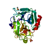
| Title | STRUCTURE OF A NOVEL EXTRACELLULAR CA2+-BINDING MODULE IN BM-40(SLASH)SPARC(SLASH)OSTEONECTIN | ||||||
 Components Components | SPARC | ||||||
 Keywords Keywords | CALCIUM-BINDING PROTEIN / EXTRACELLULAR MATRIX PROTEIN | ||||||
| Function / homology |  Function and homology information Function and homology informationsemicircular canal morphogenesis / Scavenging by Class H Receptors / platelet alpha granule membrane / platelet alpha granule / regulation of cell morphogenesis / extracellular matrix binding / negative regulation of endothelial cell proliferation / regulation of synapse organization / basement membrane / ECM proteoglycans ...semicircular canal morphogenesis / Scavenging by Class H Receptors / platelet alpha granule membrane / platelet alpha granule / regulation of cell morphogenesis / extracellular matrix binding / negative regulation of endothelial cell proliferation / regulation of synapse organization / basement membrane / ECM proteoglycans / Nuclear signaling by ERBB4 / collagen binding / endocytic vesicle lumen / positive regulation of endothelial cell migration / platelet alpha granule lumen / negative regulation of angiogenesis / nuclear matrix / Platelet degranulation / extracellular matrix / calcium ion binding / cell surface / : / extracellular region / plasma membrane / cytoplasm Similarity search - Function | ||||||
| Biological species |  Homo sapiens (human) Homo sapiens (human) | ||||||
| Method |  X-RAY DIFFRACTION / SYNCHROTRON / Resolution: 2 Å X-RAY DIFFRACTION / SYNCHROTRON / Resolution: 2 Å | ||||||
 Authors Authors | Hohenester, E. / Maurer, P. / Hohenadl, C. / Timpl, R. / Jansonius, J.N. / Engel, J. | ||||||
 Citation Citation | Journal: Nat.Struct.Biol. / Year: 1996 Title: Structure of a novel extracellular Ca(2+)-binding module in BM-40. Authors: Hohenester, E. / Maurer, P. / Hohenadl, C. / Timpl, R. / Jansonius, J.N. / Engel, J. #1: Journal: J.Mol.Biol. / Year: 1995Title: The C-Terminal Portion of Bm-40 (Sparc(Slash)Osteonectin) is an Autonomously Folding and Crystallisable Domain that Binds Calcium and Collagen Iv Authors: Maurer, P. / Hohenadl, C. / Hohenester, E. / Gohring, W. / Timpl, R. / Engel, J. #2: Journal: Faseb J. / Year: 1994Title: The Biology of Sparc, a Protein that Modulates Cell-Matrix Interactions Authors: Lane, T.F. / Sage, E.H. | ||||||
| History |
|
- Structure visualization
| Structure viewer | Molecule: MolmilJmol/JSmol |
|---|
- Downloads & links
-Download
| PDBx/mmCIF format | 1sra.cif.gz | 48.3 KB | Display | PDBx/mmCIF format |
|---|---|---|---|---|
| PDB format | pdb1sra.ent.gz | 33.3 KB | Display | PDB format |
| PDBx/mmJSON format | 1sra.json.gz | Tree view | PDBx/mmJSON format | |
| Others |  Other downloads Other downloads |
-Validation report
| Arichive directory | https://data.pdbj.org/pub/pdb/validation_reports/sr/1sraftp://data.pdbj.org/pub/pdb/validation_reports/sr/1sra | HTTPS FTP |
|---|
-Related structure data
| Similar structure data |
|---|

-Links
 PDBj
PDBj


- Assembly
| Deposited unit | 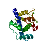
| ||||||||
|---|---|---|---|---|---|---|---|---|---|
| 1 |
| ||||||||
| Unit cell |
| ||||||||
| Atom site foot note | 1: CIS PROLINE - PRO 225 |
-Components
| #1: Protein | Mass: 17940.428 Da / Num. of mol.: 1 / Fragment: CARBOXY-TERMINAL DOMAIN (RESIDUES 136 - 286) Source method: isolated from a genetically manipulated source Details: CRYSTALLIZED FROM 0.7 M K, NA-TARTRATE, PH 7.5 + 2 MM CACL2 Source: (gene. exp.) Homo sapiens (human) / Gene: HUMAN BM-40 / Organ: KIDNEY / Plasmid: BLUESCRIPT, PCISCell line (production host): HUMAN EMBRYONIC KIDNEY CELLS (293, ATCC CRL 1573) Gene (production host): HUMAN BM-40 / Production host: Homo sapiens (human) / References: UniProt: P09486 | ||||||||
|---|---|---|---|---|---|---|---|---|---|
| #2: Chemical |   Mass: 40.078 Da / Num. of mol.: 3 / Source method: obtained synthetically / Formula: Ca Mass: 40.078 Da / Num. of mol.: 3 / Source method: obtained synthetically / Formula: Ca#3: Water | ChemComp-HOH / |  Mass: 18.015 Da / Num. of mol.: 112 / Source method: isolated from a natural source / Formula: H2O Mass: 18.015 Da / Num. of mol.: 112 / Source method: isolated from a natural source / Formula: H2OHas protein modification | Y | Nonpolymer details | CA 303 IS A NONIDENTIF | Source details | MOLECULE_NAME: SPARC. FRAGMENT 136 - 286 PRODUCED BY PCR. | |
-Experimental details
-Experiment
| Experiment | Method: X-RAY DIFFRACTION |
|---|
- Sample preparation
| Crystal | Density Matthews: 3.18 Å3/Da / Density % sol: 61.26 % | ||||||||||||||||||
|---|---|---|---|---|---|---|---|---|---|---|---|---|---|---|---|---|---|---|---|
| Crystal grow | pH: 7.5 / Details: pH 7.5 | ||||||||||||||||||
| Crystal | *PLUS Density % sol: 61 % | ||||||||||||||||||
| Crystal grow | *PLUS Method: vapor diffusion, hanging drop | ||||||||||||||||||
| Components of the solutions | *PLUS
|
-Data collection
| Diffraction source | Source: SYNCHROTRON / Site: MPG/DESY, HAMBURG  / Beamline: BW6 / Wavelength: 1.02 / Beamline: BW6 / Wavelength: 1.02 |
|---|---|
| Detector | Type: MARRESEARCH / Detector: IMAGE PLATE / Date: Apr 5, 1995 |
| Radiation | Monochromatic (M) / Laue (L): M / Scattering type: x-ray |
| Radiation wavelength | Wavelength: 1.02 Å / Relative weight: 1 |
| Reflection | Resolution: 2→20 Å / Num. obs: 15597 / % possible obs: 98.5 % / Observed criterion σ(I): 0 / Redundancy: 3.6 % / Rmerge(I) obs: 0.063 |
| Reflection | *PLUS Num. measured all: 56738 / Rmerge(I) obs: 0.063 |
| Reflection shell | *PLUS Redundancy: 3.6 % |
- Processing
| Software |
| ||||||||||||||||||||||||||||||||||||||||||||||||||||||||||||
|---|---|---|---|---|---|---|---|---|---|---|---|---|---|---|---|---|---|---|---|---|---|---|---|---|---|---|---|---|---|---|---|---|---|---|---|---|---|---|---|---|---|---|---|---|---|---|---|---|---|---|---|---|---|---|---|---|---|---|---|---|---|
| Refinement | Resolution: 2→8 Å / σ(F): 0
| ||||||||||||||||||||||||||||||||||||||||||||||||||||||||||||
| Displacement parameters | Biso mean: 24.5 Å2 | ||||||||||||||||||||||||||||||||||||||||||||||||||||||||||||
| Refine analyze | Luzzati coordinate error obs: 0.25 Å | ||||||||||||||||||||||||||||||||||||||||||||||||||||||||||||
| Refinement step | Cycle: LAST / Resolution: 2→8 Å
| ||||||||||||||||||||||||||||||||||||||||||||||||||||||||||||
| Refine LS restraints |
| ||||||||||||||||||||||||||||||||||||||||||||||||||||||||||||
| Software | *PLUS Name: X-PLOR / Classification: refinement | ||||||||||||||||||||||||||||||||||||||||||||||||||||||||||||
| Refinement | *PLUS | ||||||||||||||||||||||||||||||||||||||||||||||||||||||||||||
| Solvent computation | *PLUS | ||||||||||||||||||||||||||||||||||||||||||||||||||||||||||||
| Displacement parameters | *PLUS | ||||||||||||||||||||||||||||||||||||||||||||||||||||||||||||
| Refine LS restraints | *PLUS
|
