 Movie
Movie Controller


+ Open data

- Basic information
| Entry | Database: PDB / ID: 1bjt | ||||||
|---|---|---|---|---|---|---|---|
| Title | TOPOISOMERASE II RESIDUES 409-1201 | ||||||
 Components Components | TOPOISOMERASE II Type II topoisomerase Type II topoisomerase | ||||||
 Keywords Keywords | TOPOISOMERASE / QUATERNARY CHANGE / DNA-BINDING / DNA TOPOLOGY | ||||||
| Function / homology |  Function and homology information Function and homology informationreplication fork progression beyond termination site / DNA replication termination region / chromatin remodeling at centromere / sister chromatid segregation / regulation of mitotic recombination / resolution of meiotic recombination intermediates / SUMOylation of DNA replication proteins / synaptonemal complex / reciprocal meiotic recombination / DNA topoisomerase type II (double strand cut, ATP-hydrolyzing) activity ...replication fork progression beyond termination site / DNA replication termination region / chromatin remodeling at centromere / sister chromatid segregation / regulation of mitotic recombination / resolution of meiotic recombination intermediates / SUMOylation of DNA replication proteins / synaptonemal complex / reciprocal meiotic recombination / DNA topoisomerase type II (double strand cut, ATP-hydrolyzing) activity / DNA topoisomerase (ATP-hydrolysing) / DNA strand elongation involved in DNA replication / rRNA transcription / DNA topological change / chromatin organization / mitochondrion / DNA binding / ATP binding / identical protein binding / metal ion binding / nucleusSimilarity search - Function | ||||||
| Biological species |  Saccharomyces cerevisiae (brewer's yeast) Saccharomyces cerevisiae (brewer's yeast) | ||||||
| Method | X-RAY DIFFRACTION / SYNCHROTRON / MIR / Resolution: 2.5 Å | ||||||
 Authors Authors | Fass, D. / Bogden, C.E. / Berger, J.M. | ||||||
 Citation Citation | Journal: Nat.Struct.Biol. / Year: 1999 Title: Quaternary changes in topoisomerase II may direct orthogonal movement of two DNA strands. Authors: Fass, D. / Bogden, C.E. / Berger, J.M. #1: Journal: Nature / Year: 1996Title: Structure and Mechanism of DNA Topoisomerase II Authors: Berger, J.M. / Gamblin, S.J. / Harrison, S.C. / Wang, J.C. #2: Journal: Nature / Year: 1996Title: Erratum. Structure and Mechanism of DNA Topoisomerase II Authors: Berger, J.M. / Gamblin, S.J. / Harrison, S.C. / Wang, J.C. | ||||||
| History |
|
- Structure visualization
| Structure viewer | Molecule: MolmilJmol/JSmol |
|---|
- Downloads & links
-Download
| PDBx/mmCIF format | 1bjt.cif.gz | 158.5 KB | Display | PDBx/mmCIF format |
|---|---|---|---|---|
| PDB format | pdb1bjt.ent.gz | 121.6 KB | Display | PDB format |
| PDBx/mmJSON format | 1bjt.json.gz | Tree view | PDBx/mmJSON format | |
| Others |  Other downloads Other downloads |
-Validation report
| Arichive directory | https://data.pdbj.org/pub/pdb/validation_reports/bj/1bjtftp://data.pdbj.org/pub/pdb/validation_reports/bj/1bjt | HTTPS FTP |
|---|
-Related structure data
| Similar structure data |
|---|


-Links
 PDBj
PDBj


- Assembly
| Deposited unit | 
| ||||||||
|---|---|---|---|---|---|---|---|---|---|
| 1 | 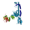
| ||||||||
| Unit cell |
|
-Components
| #1: Protein | Type II topoisomerase Mass: 91810.602 Da / Num. of mol.: 1 / Fragment: DNA-BINDING AND CLEAVAGE CORE, RESIDUES 409-1201 Source method: isolated from a genetically manipulated source Source: (gene. exp.) Saccharomyces cerevisiae (brewer's yeast)Production host: Saccharomyces cerevisiae (brewer's yeast) / References: UniProt: P06786, EC: 5.99.1.3 |
|---|---|
| #2: Water | ChemComp-HOH / Water Mass: 18.015 Da / Num. of mol.: 217 / Source method: isolated from a natural source / Formula: H2O Mass: 18.015 Da / Num. of mol.: 217 / Source method: isolated from a natural source / Formula: H2O |
-Experimental details
-Experiment
| Experiment | Method: X-RAY DIFFRACTION / Number of used crystals: 1 |
|---|
- Sample preparation
| Crystal | Density Matthews: 3.31 Å3/Da / Density % sol: 62.81 % | ||||||||||||||||||||||||||||||
|---|---|---|---|---|---|---|---|---|---|---|---|---|---|---|---|---|---|---|---|---|---|---|---|---|---|---|---|---|---|---|---|
| Crystal grow | pH: 8 / Details: pH 8.0 | ||||||||||||||||||||||||||||||
| Crystal grow | *PLUS Temperature: 4 ℃ / Method: vapor diffusion, hanging drop | ||||||||||||||||||||||||||||||
| Components of the solutions | *PLUS
|
-Data collection
| Diffraction | Mean temperature: 118 K |
|---|---|
| Diffraction source | Source: SYNCHROTRON / Site: NSLS  / Beamline: X4A / Wavelength: 1.1402 / Beamline: X4A / Wavelength: 1.1402 |
| Detector | Type: FUJI / Detector: IMAGE PLATE / Date: Mar 1, 1997 / Details: MIRRORS |
| Radiation | Monochromator: NI FILTER / Monochromatic (M) / Laue (L): M / Scattering type: x-ray |
| Radiation wavelength | Wavelength: 1.1402 Å / Relative weight: 1 |
| Reflection | Resolution: 2.5→20 Å / Num. obs: 41611 / % possible obs: 97.6 % / Observed criterion σ(I): -1.5 / Redundancy: 3.5 % / Rmerge(I) obs: 0.06 / Rsym value: 0.06 / Net I/σ(I): 29.8 |
| Reflection shell | Resolution: 2.5→2.59 Å / Redundancy: 2.7 % / Rmerge(I) obs: 0.206 / Mean I/σ(I) obs: 4.5 / Rsym value: 0.206 / % possible all: 95 |
- Processing
| Software |
| ||||||||||||||||||||||||||||||||||||||||||||||||||||||||||||
|---|---|---|---|---|---|---|---|---|---|---|---|---|---|---|---|---|---|---|---|---|---|---|---|---|---|---|---|---|---|---|---|---|---|---|---|---|---|---|---|---|---|---|---|---|---|---|---|---|---|---|---|---|---|---|---|---|---|---|---|---|---|
| Refinement | Method to determine structure: MIR / Resolution: 2.5→14 Å / Rfactor Rfree error: 0.005 / Data cutoff high absF: 10000000 / Data cutoff low absF: 0.001 / Cross valid method: THROUGHOUT / σ(F): 2 Details: THE B FACTORS FOR RESIDUES 653 - 659 ARE VERY HIGH, BUT THE DENSITY FOR THESE RESIDUES WAS SEEN IN SOLVENT-FLATTENED MIR MAPS. THESE RESIDUES HELPED ASSIGN THE CONNECTIVITY BETWEEN DOMAINS IN THE DIMER.
| ||||||||||||||||||||||||||||||||||||||||||||||||||||||||||||
| Refinement step | Cycle: LAST / Resolution: 2.5→14 Å
| ||||||||||||||||||||||||||||||||||||||||||||||||||||||||||||
| Refine LS restraints |
| ||||||||||||||||||||||||||||||||||||||||||||||||||||||||||||
| LS refinement shell | Resolution: 2.5→2.61 Å / Rfactor Rfree error: 0.019 / Total num. of bins used: 8
| ||||||||||||||||||||||||||||||||||||||||||||||||||||||||||||
| Software | *PLUS Name: X-PLOR / Version: 3.851 / Classification: refinement | ||||||||||||||||||||||||||||||||||||||||||||||||||||||||||||
| Refine LS restraints | *PLUS
|
