 Movie
Movie Controller

[English] 日本語
 Yorodumi
- PDB-5fb3: Structure of glycerophosphate dehydrogenase in complex with NADPH -
+ Open data

- Basic information
| Entry | Database: PDB / ID: 5fb3 | ||||||
|---|---|---|---|---|---|---|---|
| Title | Structure of glycerophosphate dehydrogenase in complex with NADPH | ||||||
 Components Components | Glycerol-1-phosphate dehydrogenase [NAD(P)+] | ||||||
 Keywords Keywords | OXIDOREDUCTASE / Rossmann fold | ||||||
| Function / homology |  Function and homology information Function and homology informationsn-glycerol-1-phosphate dehydrogenase / glycerol-1-phosphate dehydrogenase (NAD+) activity / glycerol-1-phosphate dehydrogenase (NADP+) activity / glycerophospholipid metabolic process / phospholipid biosynthetic process / metal ion binding / cytoplasm Similarity search - Function | ||||||
| Biological species |   Pyrobaculum calidifontis JCM 11548 (archaea) Pyrobaculum calidifontis JCM 11548 (archaea) | ||||||
| Method |  X-RAY DIFFRACTION / SYNCHROTRON / SIRAS / Resolution: 2.45 Å X-RAY DIFFRACTION / SYNCHROTRON / SIRAS / Resolution: 2.45 Å | ||||||
 Authors Authors | Sakuraba, H. / Hayashi, J. / Yamamoto, K. / Yoneda, K. / Ohshima, T. | ||||||
 Citation Citation | Journal: Proteins / Year: 2016 Title: Unique coenzyme binding mode of hyperthermophilic archaeal sn-glycerol-1-phosphate dehydrogenase from Pyrobaculum calidifontis Authors: Hayashi, J. / Yamamoto, K. / Yoneda, K. / Ohshima, T. / Sakuraba, H. | ||||||
| History |
|
- Structure visualization
| Structure viewer | Molecule: MolmilJmol/JSmol |
|---|
- Downloads & links
-Download
| PDBx/mmCIF format | 5fb3.cif.gz | 392.3 KB | Display | PDBx/mmCIF format |
|---|---|---|---|---|
| PDB format | pdb5fb3.ent.gz | 319.7 KB | Display | PDB format |
| PDBx/mmJSON format | 5fb3.json.gz | Tree view | PDBx/mmJSON format | |
| Others |  Other downloads Other downloads |
-Validation report
| Arichive directory | https://data.pdbj.org/pub/pdb/validation_reports/fb/5fb3ftp://data.pdbj.org/pub/pdb/validation_reports/fb/5fb3 | HTTPS FTP |
|---|
-Related structure data
| Similar structure data |
|---|


-Links
 PDBj
PDBj

- Assembly
| Deposited unit | 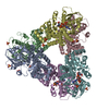
| ||||||||
|---|---|---|---|---|---|---|---|---|---|
| 1 |
| ||||||||
| Unit cell |
|
-Components
-Protein , 1 types, 6 molecules ABCDEF
| #1: Protein | Mass: 37042.953 Da / Num. of mol.: 6 Source method: isolated from a genetically manipulated source Source: (gene. exp.) Pyrobaculum calidifontis JCM 11548 (archaea)Strain: JCM 11548 / Gene: egsA, Pcal_0566 / Plasmid: pET11a / Production host:  References: UniProt: A3MTM6, sn-glycerol-1-phosphate dehydrogenase |
|---|
-Non-polymers , 5 types, 187 molecules 


| #2: Chemical | ChemComp-ZN /  Mass: 65.409 Da / Num. of mol.: 6 / Source method: obtained synthetically / Formula: Zn Mass: 65.409 Da / Num. of mol.: 6 / Source method: obtained synthetically / Formula: Zn#3: Chemical | ChemComp-SO4 /  Mass: 96.063 Da / Num. of mol.: 12 / Source method: obtained synthetically / Formula: SO4 Mass: 96.063 Da / Num. of mol.: 12 / Source method: obtained synthetically / Formula: SO4#4: Chemical | ChemComp-PPV / |  Mass: 177.975 Da / Num. of mol.: 1 / Source method: obtained synthetically / Formula: H4O7P2 Mass: 177.975 Da / Num. of mol.: 1 / Source method: obtained synthetically / Formula: H4O7P2#5: Chemical |  Mass: 745.421 Da / Num. of mol.: 3 / Source method: obtained synthetically / Formula: C21H30N7O17P3 Mass: 745.421 Da / Num. of mol.: 3 / Source method: obtained synthetically / Formula: C21H30N7O17P3#6: Water | ChemComp-HOH / | Mass: 18.015 Da / Num. of mol.: 165 / Source method: isolated from a natural source / Formula: H2O |
|---|
-Experimental details
-Experiment
| Experiment | Method: X-RAY DIFFRACTION / Number of used crystals: 1 |
|---|
- Sample preparation
| Crystal | Density Matthews: 2.36 Å3/Da / Density % sol: 47.96 % |
|---|---|
| Crystal grow | Temperature: 293 K / Method: vapor diffusion, sitting drop / pH: 6.5 / Details: ammonium sulfate |
-Data collection
| Diffraction | Mean temperature: 100 K | |||||||||||||||||||||||||||||||||||||||||||||||||||||||||||||||||||||||||||||||||||||||||||||||||||||||||||||||||||||||||||||||||||||||||||||||||||||||||||||||||||||||||||||||||||||||||||||
|---|---|---|---|---|---|---|---|---|---|---|---|---|---|---|---|---|---|---|---|---|---|---|---|---|---|---|---|---|---|---|---|---|---|---|---|---|---|---|---|---|---|---|---|---|---|---|---|---|---|---|---|---|---|---|---|---|---|---|---|---|---|---|---|---|---|---|---|---|---|---|---|---|---|---|---|---|---|---|---|---|---|---|---|---|---|---|---|---|---|---|---|---|---|---|---|---|---|---|---|---|---|---|---|---|---|---|---|---|---|---|---|---|---|---|---|---|---|---|---|---|---|---|---|---|---|---|---|---|---|---|---|---|---|---|---|---|---|---|---|---|---|---|---|---|---|---|---|---|---|---|---|---|---|---|---|---|---|---|---|---|---|---|---|---|---|---|---|---|---|---|---|---|---|---|---|---|---|---|---|---|---|---|---|---|---|---|---|---|---|---|
| Diffraction source | Source: SYNCHROTRON / Site: Photon Factory  / Beamline: AR-NW12A / Wavelength: 1 Å / Beamline: AR-NW12A / Wavelength: 1 Å | |||||||||||||||||||||||||||||||||||||||||||||||||||||||||||||||||||||||||||||||||||||||||||||||||||||||||||||||||||||||||||||||||||||||||||||||||||||||||||||||||||||||||||||||||||||||||||||
| Detector | Type: ADSC QUANTUM 270 / Detector: CCD / Date: Jun 12, 2015 | |||||||||||||||||||||||||||||||||||||||||||||||||||||||||||||||||||||||||||||||||||||||||||||||||||||||||||||||||||||||||||||||||||||||||||||||||||||||||||||||||||||||||||||||||||||||||||||
| Radiation | Monochromator: Si / Protocol: SINGLE WAVELENGTH / Monochromatic (M) / Laue (L): M / Scattering type: x-ray | |||||||||||||||||||||||||||||||||||||||||||||||||||||||||||||||||||||||||||||||||||||||||||||||||||||||||||||||||||||||||||||||||||||||||||||||||||||||||||||||||||||||||||||||||||||||||||||
| Radiation wavelength | Wavelength: 1 Å / Relative weight: 1 | |||||||||||||||||||||||||||||||||||||||||||||||||||||||||||||||||||||||||||||||||||||||||||||||||||||||||||||||||||||||||||||||||||||||||||||||||||||||||||||||||||||||||||||||||||||||||||||
| Reflection | Resolution: 2.45→50 Å / Num. obs: 78117 / % possible obs: 100 % / Redundancy: 6.7 % / Rmerge(I) obs: 0.07 / Rpim(I) all: 0.029 / Rrim(I) all: 0.076 / Χ2: 0.75 / Net I/av σ(I): 25.771 / Net I/σ(I): 7.2 / Num. measured all: 520646 | |||||||||||||||||||||||||||||||||||||||||||||||||||||||||||||||||||||||||||||||||||||||||||||||||||||||||||||||||||||||||||||||||||||||||||||||||||||||||||||||||||||||||||||||||||||||||||||
| Reflection shell | Diffraction-ID: 1 / Rejects: _
|
- Processing
| Software |
| |||||||||||||||||||||||||||||||||||||||||||||||||||||||||||||||||||||||||||
|---|---|---|---|---|---|---|---|---|---|---|---|---|---|---|---|---|---|---|---|---|---|---|---|---|---|---|---|---|---|---|---|---|---|---|---|---|---|---|---|---|---|---|---|---|---|---|---|---|---|---|---|---|---|---|---|---|---|---|---|---|---|---|---|---|---|---|---|---|---|---|---|---|---|---|---|---|
| Refinement | Method to determine structure: SIRAS / Resolution: 2.45→50 Å / Cor.coef. Fo:Fc: 0.952 / Cor.coef. Fo:Fc free: 0.931 / SU B: 11.703 / SU ML: 0.26 / Cross valid method: THROUGHOUT / σ(F): 0 / ESU R: 0.63 / ESU R Free: 0.3 / Stereochemistry target values: MAXIMUM LIKELIHOOD Details: HYDROGENS HAVE BEEN ADDED IN THE RIDING POSITIONS U VALUES : REFINED INDIVIDUALLY
| |||||||||||||||||||||||||||||||||||||||||||||||||||||||||||||||||||||||||||
| Solvent computation | Ion probe radii: 0.8 Å / Shrinkage radii: 0.8 Å / VDW probe radii: 1.2 Å / Solvent model: MASK | |||||||||||||||||||||||||||||||||||||||||||||||||||||||||||||||||||||||||||
| Displacement parameters | Biso max: 146.28 Å2 / Biso mean: 63.236 Å2 / Biso min: 20 Å2
| |||||||||||||||||||||||||||||||||||||||||||||||||||||||||||||||||||||||||||
| Refinement step | Cycle: final / Resolution: 2.45→50 Å
| |||||||||||||||||||||||||||||||||||||||||||||||||||||||||||||||||||||||||||
| Refine LS restraints |
| |||||||||||||||||||||||||||||||||||||||||||||||||||||||||||||||||||||||||||
| LS refinement shell | Resolution: 2.447→2.51 Å / Total num. of bins used: 20
|
