 Movie
Movie Controller


+ Open data

- Basic information
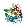
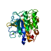
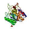
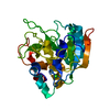
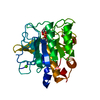
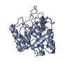
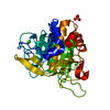
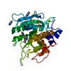
| Entry | Database: PDB / ID: 1aqn | ||||||
|---|---|---|---|---|---|---|---|
| Title | SUBTILISIN MUTANT 8324 | ||||||
 Components Components | SUBTILISIN 8324 | ||||||
 Keywords Keywords | SERINE PROTEASE / HYDROLASE / SERINE PROTEINASE | ||||||
| Function / homology |  Function and homology information Function and homology informationsubtilisin / sporulation resulting in formation of a cellular spore / fibrinolysis / serine-type endopeptidase activity / proteolysis / extracellular region / metal ion binding Similarity search - Function | ||||||
| Biological species |  | ||||||
| Method |  X-RAY DIFFRACTION / DIFFERENCE MAP / Resolution: 1.8 Å X-RAY DIFFRACTION / DIFFERENCE MAP / Resolution: 1.8 Å | ||||||
 Authors Authors | Whitlow, M. / Howard, A.J. / Wood, J.F. | ||||||
 Citation Citation | Journal: To be published Title: Large increases in general stabilityfor subtilisin BPN' through incremental changes in the free energy of unfolding Authors: Muralikrishna, P. / Wickstrom, E. #1: Journal: Biochemistry / Year: 1988Title: The Engineering of Binding Affinity at Metal Ion Binding Sites for the Stabilization of Proteins: Subtilisin as a Test Case Authors: Pantoliano, M.W. / Whitlow, M. / Wood, J.F. / Rollence, M.L. / Finzel, B.C. / Gilliland, G.L. / Poulos, T.L. / Bryan, P.N. #2: Journal: Biochemistry / Year: 1987Title: Protein Engineering of Subtilisin Bpn': Enhanced Stabilization Through the Introduction of Two Cysteines to Form a Disulfide Bond Authors: Pantoliano, M.W. / Ladner, R.C. / Bryan, P.N. / Rollence, M.L. / Wood, J.F. / Poulos, T.L. #3: Journal: Proteins / Year: 1986Title: Proteases of Enhanced Stability: Characterization of a Thermostable Variant of Subtilisin Authors: Bryan, P.N. / Rollence, M.L. / Pantoliano, M.W. / Wood, J. / Finzel, B.C. / Gilliland, G.L. / Howard, A.J. / Poulos, T.L. #4: Journal: Biochem.Biophys.Res.Commun. / Year: 1971Title: Atomic Coordinates for Subtilisin Bpn' (or Novo) Authors: Alden, R.A. / Birktoft, J.J. / Kraut, J. / Robertus, J.D. / Wright, C.S. | ||||||
| History |
|
- Structure visualization
| Structure viewer | Molecule: MolmilJmol/JSmol |
|---|
- Downloads & links
-Download
| PDBx/mmCIF format | 1aqn.cif.gz | 68.3 KB | Display | PDBx/mmCIF format |
|---|---|---|---|---|
| PDB format | pdb1aqn.ent.gz | 49 KB | Display | PDB format |
| PDBx/mmJSON format | 1aqn.json.gz | Tree view | PDBx/mmJSON format | |
| Others |  Other downloads Other downloads |
-Validation report
| Arichive directory | https://data.pdbj.org/pub/pdb/validation_reports/aq/1aqnftp://data.pdbj.org/pub/pdb/validation_reports/aq/1aqn | HTTPS FTP |
|---|
-Related structure data
| Similar structure data |
|---|
-Links
 PDBj
PDBj


- Assembly
| Deposited unit | 
| ||||||||
|---|---|---|---|---|---|---|---|---|---|
| 1 |
| ||||||||
| Unit cell |
|
-Components
| #1: Protein | Mass: 27514.631 Da / Num. of mol.: 1 / Mutation: T22C, M50F, S87C, G169A, Q206C, Y217K, N218S Source method: isolated from a genetically manipulated source Source: (gene. exp.) | ||||||||
|---|---|---|---|---|---|---|---|---|---|
| #2: Chemical |   Mass: 40.078 Da / Num. of mol.: 2 / Source method: obtained synthetically / Formula: Ca Mass: 40.078 Da / Num. of mol.: 2 / Source method: obtained synthetically / Formula: Ca#3: Chemical |   Num. of mol.: 2 / Source method: obtained synthetically Num. of mol.: 2 / Source method: obtained synthetically#4: Chemical |   Mass: 60.095 Da / Num. of mol.: 2 / Source method: obtained synthetically / Formula: C3H8O / Comment: alkaloid*YM Mass: 60.095 Da / Num. of mol.: 2 / Source method: obtained synthetically / Formula: C3H8O / Comment: alkaloid*YM#5: Water | ChemComp-HOH / |  Mass: 18.015 Da / Num. of mol.: 198 / Source method: isolated from a natural source / Formula: H2O Mass: 18.015 Da / Num. of mol.: 198 / Source method: isolated from a natural source / Formula: H2OHas protein modification | Y | |
-Experimental details
-Experiment
| Experiment | Method: X-RAY DIFFRACTION / Number of used crystals: 1 |
|---|
- Sample preparation
| Crystal | Density Matthews: 2.04 Å3/Da / Density % sol: 39.61 % |
|---|---|
| Crystal grow | Method: vapor diffusion / pH: 8.7 Details: CRYSTAL WERE GROWN BY VAPOR DIFFUSION OF 10 MG/ML PROTEIN IN 100 MM TRIS-HCL PH 8.7, 40 MM CACL2 AGAINST 20% 2-PROPANOL., vapor diffusion |
| Crystal grow | *PLUS Method: vapor diffusion, hanging dropDetails: Bryan, P.N., (1986) Proteins: Struct.,Funct., Genet., 1, 326. |
| Components of the solutions | *PLUS Conc.: 55 % / Common name: acetone |
-Data collection
| Diffraction | Mean temperature: 290 K |
|---|---|
| Diffraction source | Source: ROTATING ANODE / Type: ELLIOTT GX-21 / Wavelength: 1.5418 |
| Detector | Type: SIEMENS / Detector: AREA DETECTOR / Date: May 20, 1987 / Details: HUBER MONOCHROMATOR |
| Radiation | Monochromatic (M) / Laue (L): M / Scattering type: x-ray |
| Radiation wavelength | Wavelength: 1.5418 Å / Relative weight: 1 |
| Reflection | Resolution: 1.73→99 Å / Num. obs: 18552 / % possible obs: 79.8 % / Observed criterion σ(I): 2 / Redundancy: 2.7 % / Rsym value: 0.0634 / Net I/σ(I): 17.3 |
| Reflection shell | Resolution: 1.73→1.84 Å / Redundancy: 2.33 % / Mean I/σ(I) obs: 2.9 / Rsym value: 0.3174 / % possible all: 41.4 |
- Processing
| Software |
| ||||||||||||||||||||||||||||||||||||||||||||||||||||||||||||||||||||||||||||||||||||
|---|---|---|---|---|---|---|---|---|---|---|---|---|---|---|---|---|---|---|---|---|---|---|---|---|---|---|---|---|---|---|---|---|---|---|---|---|---|---|---|---|---|---|---|---|---|---|---|---|---|---|---|---|---|---|---|---|---|---|---|---|---|---|---|---|---|---|---|---|---|---|---|---|---|---|---|---|---|---|---|---|---|---|---|---|---|
| Refinement | Method to determine structure: DIFFERENCE MAP / Resolution: 1.8→10 Å / Num. reflection all: 16735 / Num. reflection obs: 16735 / σ(F): 2 | ||||||||||||||||||||||||||||||||||||||||||||||||||||||||||||||||||||||||||||||||||||
| Refinement step | Cycle: LAST / Resolution: 1.8→10 Å
| ||||||||||||||||||||||||||||||||||||||||||||||||||||||||||||||||||||||||||||||||||||
| Refine LS restraints |
| ||||||||||||||||||||||||||||||||||||||||||||||||||||||||||||||||||||||||||||||||||||
| Software | *PLUS Name: PROFFT / Classification: refinement | ||||||||||||||||||||||||||||||||||||||||||||||||||||||||||||||||||||||||||||||||||||
| Refinement | *PLUS Rfactor obs: 0.139 / Rfactor Rwork: 0.139 | ||||||||||||||||||||||||||||||||||||||||||||||||||||||||||||||||||||||||||||||||||||
| Solvent computation | *PLUS | ||||||||||||||||||||||||||||||||||||||||||||||||||||||||||||||||||||||||||||||||||||
| Displacement parameters | *PLUS |
