 Movie
Movie Controller


+ Open data

- Basic information
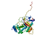
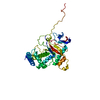
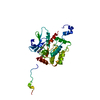
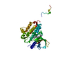
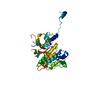
| Entry | Database: PDB / ID: 1anv | ||||||
|---|---|---|---|---|---|---|---|
| Title | ADENOVIRUS 5 DBP/URANYL FLUORIDE SOAK | ||||||
 Components Components | ADENOVIRUS SINGLE-STRANDED DNA-BINDING PROTEIN | ||||||
 Keywords Keywords |  DNA BINDING PROTEIN / EARLY PROTEIN / DNA-BINDING PROTEIN / ZINC-FINGER / PHOSPHORYLATION / NUCLEAR PROTEIN DNA BINDING PROTEIN / EARLY PROTEIN / DNA-BINDING PROTEIN / ZINC-FINGER / PHOSPHORYLATION / NUCLEAR PROTEIN | ||||||
| Function / homology |  Function and homology information Function and homology informationviral DNA strand displacement replication / viral DNA genome replication / DNA duplex unwinding / DNA unwinding involved in DNA replication / positive regulation of DNA replication / viral capsid / single-stranded DNA binding / DNA-templated transcription / host cell nucleus / DNA binding ...viral DNA strand displacement replication / viral DNA genome replication / DNA duplex unwinding / DNA unwinding involved in DNA replication / positive regulation of DNA replication / viral capsid / single-stranded DNA binding / DNA-templated transcription / host cell nucleus / DNA binding / zinc ion binding / identical protein bindingSimilarity search - Function | ||||||
| Biological species |   Human adenovirus 5 Human adenovirus 5 | ||||||
| Method | X-RAY DIFFRACTION / MOLECULAR REPLACEMENT / Resolution: 2.7 Å | ||||||
 Authors Authors | Kanellopoulos, P.N. / Tsernoglou, D. / Van Der Vliet, P.C. / Tucker, P.A. | ||||||
 Citation Citation | Journal: Acta Crystallogr.,Sect.D / Year: 1996 Title: Conformational change of the adenovirus DNA-binding protein induced by soaking crystals with K3UO2F5 solutions. Authors: Kanellopoulos, P.N. / Tsernoglou, D. / van der Vliet, P.C. / Tucker, P.A. #1: Journal: J.Mol.Biol. / Year: 1996Title: Alternative Arrangements of the Protein Chain are Possible for the Adenovirus Single-Stranded DNA Binding Protein Authors: Kanellopoulos, P.N. / Tsernoglou, D. / Van Der Vliet, P.C. / Tucker, P.A. #2: Journal: Embo J. / Year: 1994Title: Crystal Structure of the Adenovirus DNA Binding Protein Reveals a Hook-on Model for Cooperative DNA Binding Authors: Tucker, P.A. / Tsernoglou, D. / Tucker, A.D. / Coenjaerts, F.E. / Leenders, H. / Van Der Vliet, P.C. | ||||||
| History |
|
- Structure visualization
| Structure viewer | Molecule: MolmilJmol/JSmol |
|---|
- Downloads & links
-Download
| PDBx/mmCIF format | 1anv.cif.gz | 72.8 KB | Display | PDBx/mmCIF format |
|---|---|---|---|---|
| PDB format | pdb1anv.ent.gz | 56.5 KB | Display | PDB format |
| PDBx/mmJSON format | 1anv.json.gz | Tree view | PDBx/mmJSON format | |
| Others |  Other downloads Other downloads |
-Validation report
| Arichive directory | https://data.pdbj.org/pub/pdb/validation_reports/an/1anvftp://data.pdbj.org/pub/pdb/validation_reports/an/1anv | HTTPS FTP |
|---|
-Related structure data
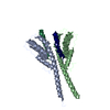
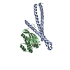
| Related structure data |  1adt S: Starting model for refinement |
|---|---|
| Similar structure data |
-Links
 PDBj
PDBj
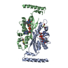
- Assembly
| Deposited unit | 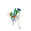
| ||||||||
|---|---|---|---|---|---|---|---|---|---|
| 1 |
| ||||||||
| Unit cell |
|
-Components
| #1: Protein | Mass: 39899.746 Da / Num. of mol.: 1 / Fragment: C-TERMINAL DOMAIN, RESIDUES 174 - 529 / Source method: isolated from a natural source / Source: (natural) Human adenovirus 5 / Genus: Mastadenovirus / Species: Human adenovirus C / References: UniProt: P03265 | ||||
|---|---|---|---|---|---|
| #2: Chemical |   Mass: 65.409 Da / Num. of mol.: 2 / Source method: obtained synthetically / Formula: Zn Mass: 65.409 Da / Num. of mol.: 2 / Source method: obtained synthetically / Formula: Zn#3: Chemical | ChemComp-IUM / Uranyl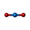  Mass: 270.028 Da / Num. of mol.: 4 / Source method: obtained synthetically / Formula: O2U Mass: 270.028 Da / Num. of mol.: 4 / Source method: obtained synthetically / Formula: O2U#4: Water | ChemComp-HOH / | Water Mass: 18.015 Da / Num. of mol.: 31 / Source method: isolated from a natural source / Formula: H2O Mass: 18.015 Da / Num. of mol.: 31 / Source method: isolated from a natural source / Formula: H2O |
-Experimental details
-Experiment
| Experiment | Method: X-RAY DIFFRACTION / Number of used crystals: 1 |
|---|
- Sample preparation
| Crystal | Density Matthews: 2.29 Å3/Da / Density % sol: 46.21 % | |||||||||||||||
|---|---|---|---|---|---|---|---|---|---|---|---|---|---|---|---|---|
| Crystal grow | pH: 5.8 / Details: pH 5.8 | |||||||||||||||
| Crystal grow | *PLUS Method: vapor diffusion / Details: Tsernoglou, D., (1984) J. Mol. Biol., 172, 237. | |||||||||||||||
| Components of the solutions | *PLUS
|
-Data collection
| Diffraction | Mean temperature: 295 K |
|---|---|
| Diffraction source | Source: ROTATING ANODE / Type: MACSCIENCE M18X / Wavelength: 1.5418 |
| Detector | Type: MARRESEARCH / Detector: IMAGE PLATE / Date: May 27, 1994 |
| Radiation | Monochromator: GRAPHITE(002) / Monochromatic (M) / Laue (L): M / Scattering type: x-ray |
| Radiation wavelength | Wavelength: 1.5418 Å / Relative weight: 1 |
| Reflection | Resolution: 2.7→15 Å / Num. obs: 10312 / % possible obs: 98.7 % / Observed criterion σ(I): 0 / Redundancy: 3.4 % / Rmerge(I) obs: 0.147 |
| Reflection shell | Resolution: 2.7→3.2 Å / Redundancy: 3.4 % / Rmerge(I) obs: 0.342 / % possible all: 99.7 |
| Reflection | *PLUS Num. measured all: 34497 |
- Processing
| Software |
| ||||||||||||||||||||||||||||||
|---|---|---|---|---|---|---|---|---|---|---|---|---|---|---|---|---|---|---|---|---|---|---|---|---|---|---|---|---|---|---|---|
| Refinement | Method to determine structure: MOLECULAR REPLACEMENT Starting model: 1ADT 1adt Resolution: 2.7→15 Å / σ(F): 0
| ||||||||||||||||||||||||||||||
| Refinement step | Cycle: LAST / Resolution: 2.7→15 Å
| ||||||||||||||||||||||||||||||
| Refine LS restraints |
| ||||||||||||||||||||||||||||||
| Software | *PLUS Name: TNT / Classification: refinement | ||||||||||||||||||||||||||||||
| Refinement | *PLUS Rfactor obs: 0.206 / Rfactor Rfree: 0.314 | ||||||||||||||||||||||||||||||
| Solvent computation | *PLUS | ||||||||||||||||||||||||||||||
| Displacement parameters | *PLUS | ||||||||||||||||||||||||||||||
| Refine LS restraints | *PLUS
|
