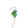
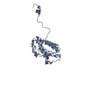
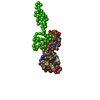
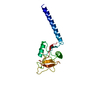
- PDB-2xze: Structural basis for AMSH-ESCRT-III CHMP3 interaction -
+
Open data
ID or keywords:
Loading...
-
Basic information
Entry
Database: PDB / ID: 2xze
Title
Structural basis for AMSH-ESCRT-III CHMP3 interaction
Components
CHARGED MULTIVESICULAR BODY PROTEIN 3
STAM-BINDING PROTEIN
Keywords
HYDROLASE/PROTEIN TRANSPORT / HYDROLASE-PROTEIN TRANSPORT COMPLEX
Function / homology
Function and homology information
regulation of endosome size / Hydrolases; Acting on peptide bonds (peptidases); Omega peptidases / negative regulation of hippocampal neuron apoptotic process / multivesicular body-lysosome fusion / amphisome membrane / vesicle fusion with vacuole / late endosome to lysosome transport / ESCRT III complex / kinetochore microtubule / endosome transport via multivesicular body sorting pathway ...regulation of endosome size / Hydrolases; Acting on peptide bonds (peptidases); Omega peptidases / negative regulation of hippocampal neuron apoptotic process / multivesicular body-lysosome fusion / amphisome membrane / vesicle fusion with vacuole / late endosome to lysosome transport / ESCRT III complex / kinetochore microtubule / endosome transport via multivesicular body sorting pathway / nuclear membrane reassembly / multivesicular body sorting pathway / midbody abscission / Sealing of the nuclear envelope (NE) by ESCRT-III / deubiquitinase activity / regulation of centrosome duplication / suppression of viral release by host / membrane fission / multivesicular body assembly / late endosome to vacuole transport / multivesicular body membrane / phosphatidylcholine binding / negative regulation of Ras protein signal transduction / ubiquitin-dependent protein catabolic process via the multivesicular body sorting pathway / plasma membrane repair / regulation of mitotic spindle assembly / Translation of Replicase and Assembly of the Replication Transcription Complex / hippocampal neuron apoptotic process / molecular function inhibitor activity / regulation of early endosome to late endosome transport / protein K63-linked deubiquitination / metal-dependent deubiquitinase activity / mitotic metaphase chromosome alignment / nucleus organization / K63-linked deubiquitinase activity / Macroautophagy / positive regulation of cytokinesis / viral budding via host ESCRT complex / ubiquitin-specific protease binding / cleavage furrow / mitotic cytokinesis / autophagosome membrane / viral release from host cell / protein polymerization / Pyroptosis / autophagosome maturation / protein deubiquitination / nuclear pore / cell surface receptor signaling pathway via JAK-STAT / multivesicular body / phosphatidylinositol-4,5-bisphosphate binding / Endosomal Sorting Complex Required For Transport (ESCRT) / viral budding from plasma membrane / negative regulation of phosphatidylinositol 3-kinase/protein kinase B signal transduction / HCMV Late Events / macroautophagy / Late endosomal microautophagy / Budding and maturation of HIV virion / kinetochore / autophagy / Metalloprotease DUBs / late endosome / protein transport / cytoplasmic vesicle / Translation of Replicase and Assembly of the Replication Transcription Complex / midbody / early endosome / endosome / protein domain specific binding / lysosomal membrane / apoptotic process / positive regulation of cell population proliferation / proteolysis / extracellular exosome / nucleoplasm / metal ion binding / identical protein binding / nucleus / plasma membrane / cytosol Similarity search - Function
Mass: 18.015 Da / Num. of mol.: 386 / Source method: isolated from a natural source / Formula: H2O
-
Experimental details
-
Experiment
Experiment
Method: X-RAY DIFFRACTION / Number of used crystals: 1
-
Sample preparation
Crystal
Density Matthews: 2.7 Å3/Da / Density % sol: 54 % / Description: NONE
Crystal grow
pH: 6 Details: 3.3 MG/ML OF AMSH PROTEIN IN 10 MM HEPES PH 8.0, 100 MM NACL WAS MIXED WITH AN EQUAL VOLUME WELL CONDITION: 2.2 M SODIUM MALONATE,WITH 30 % GLYCEROL AS CRYO-PROTECTANT
Monochromator: U35 UNDULATOR / Protocol: SINGLE WAVELENGTH / Monochromatic (M) / Laue (L): M / Scattering type: x-ray
Radiation wavelength
ID
Wavelength (Å)
Relative weight
1
0.976
1
2
0.9795
1
Reflection
Resolution: 1.75→44.85 Å / Num. obs: 141404 / % possible obs: 99.8 % / Observed criterion σ(I): 0 / Redundancy: 3.3 % / Rmerge(I) obs: 0.08 / Net I/σ(I): 5.1
Reflection shell
Resolution: 1.75→1.84 Å / Redundancy: 3.3 % / Rmerge(I) obs: 0.64 / Mean I/σ(I) obs: 1.2 / % possible all: 100
-
Processing
Software
Name
Version
Classification
REFMAC
5.2.0019
refinement
MOSFLM
datareduction
SCALA
datascaling
Auto-Rickshaw
phasing
Refinement
Method to determine structure: SAD Starting model: NONE Resolution: 1.75→51.71 Å / Cor.coef. Fo:Fc: 0.961 / Cor.coef. Fo:Fc free: 0.945 / SU B: 2.52 / SU ML: 0.08 / Cross valid method: THROUGHOUT / ESU R: 0.113 / ESU R Free: 0.111 / Stereochemistry target values: MAXIMUM LIKELIHOOD / Details: HYDROGENS HAVE BEEN ADDED IN THE RIDING POSITIONS.
Rfactor
Num. reflection
% reflection
Selection details
Rfree
0.22694
2164
5 %
RANDOM
Rwork
0.19163
-
-
-
obs
0.19333
40725
99.73 %
-
Solvent computation
Ion probe radii: 0.8 Å / Shrinkage radii: 0.8 Å / VDW probe radii: 1.2 Å / Solvent model: MASK
 Movie
Movie Controller


 Open data
 Basic information
 Components
 Keywords
 Function and homology information
 HOMO SAPIENS (human)
 X-RAY DIFFRACTION /
  Authors
 Citation
 Structure visualization
 Downloads & links
 Other downloads
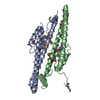


 PDBj
PDBj


 Assembly


 Mass: 18.015 Da / Num. of mol.: 386 / Source method: isolated from a natural source / Formula: H2O
 Sample preparation
 / Beamline: ID14-4 / Wavelength: 0.9760, 0.9795
 Processing