 Movie
Movie Controller

[English] 日本語
 Yorodumi
- PDB-2wb0: 2.1 resolution structure of the C-terminal domain of the human ad... -
+ Open data

- Basic information
| Entry | Database: PDB / ID: 2wb0 | |||||||||
|---|---|---|---|---|---|---|---|---|---|---|
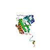
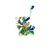
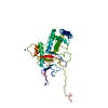
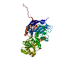
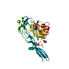
| Title | 2.1 resolution structure of the C-terminal domain of the human adenovirus 5 ssDNA binding protein | |||||||||
 Components Components | E2A DNA-BINDING PROTEIN | |||||||||
 Keywords Keywords | DNA BINDING PROTEIN / DNA-BINDING PROTEIN / SSDNA BINDING PROTEIN | |||||||||
| Function / homology |  Function and homology information Function and homology informationviral DNA strand displacement replication / viral DNA genome replication / positive regulation of DNA replication / viral capsid / single-stranded DNA binding / DNA replication / DNA-templated transcription / host cell nucleus / DNA binding / zinc ion binding / identical protein binding Similarity search - Function | |||||||||
| Biological species |   HUMAN ADENOVIRUS 5 HUMAN ADENOVIRUS 5 | |||||||||
| Method |  X-RAY DIFFRACTION / SYNCHROTRON / MOLECULAR REPLACEMENT / Resolution: 1.95 Å X-RAY DIFFRACTION / SYNCHROTRON / MOLECULAR REPLACEMENT / Resolution: 1.95 Å | |||||||||
 Authors Authors | Hendle, J. / Kanellopoulos, P.N. / Tucker, P.A. | |||||||||
 Citation Citation | Journal: To be Published Title: High Resolution Structures of the Adenovirus Single-Stranded DNA Binding Protein and the N512P Hinge-Region Mutant Authors: Hendle, J. / Kanellopoulos, P.N. / Tucker, P.A. | |||||||||
| History |
|
- Structure visualization
| Structure viewer | Molecule: MolmilJmol/JSmol |
|---|
- Downloads & links
-Download
| PDBx/mmCIF format | 2wb0.cif.gz | 86.6 KB | Display | PDBx/mmCIF format |
|---|---|---|---|---|
| PDB format | pdb2wb0.ent.gz | 63.6 KB | Display | PDB format |
| PDBx/mmJSON format | 2wb0.json.gz | Tree view | PDBx/mmJSON format | |
| Others |  Other downloads Other downloads |
-Validation report
| Arichive directory | https://data.pdbj.org/pub/pdb/validation_reports/wb/2wb0ftp://data.pdbj.org/pub/pdb/validation_reports/wb/2wb0 | HTTPS FTP |
|---|
-Related structure data
| Related structure data | 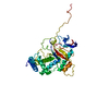 2wazC  1adt C: citing same article ( S: Starting model for refinement |
|---|---|
| Similar structure data |


-Links
 PDBj
PDBj- Assembly
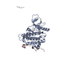
| Deposited unit | 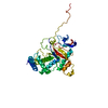
| ||||||||
|---|---|---|---|---|---|---|---|---|---|
| 1 |
| ||||||||
| Unit cell |
|
-Components
| #1: Protein | Mass: 39899.746 Da / Num. of mol.: 1 / Fragment: C-TERMINAL DNA BINDING DOMAIN, RESIDUES 174-529 Source method: isolated from a genetically manipulated source Source: (gene. exp.) HUMAN ADENOVIRUS 5 / Plasmid: PETM11 / Production host:  | ||
|---|---|---|---|
| #2: Chemical |   Mass: 65.409 Da / Num. of mol.: 2 / Source method: obtained synthetically / Formula: Zn Mass: 65.409 Da / Num. of mol.: 2 / Source method: obtained synthetically / Formula: Zn#3: Water | ChemComp-HOH / |  Mass: 18.015 Da / Num. of mol.: 383 / Source method: isolated from a natural source / Formula: H2O Mass: 18.015 Da / Num. of mol.: 383 / Source method: isolated from a natural source / Formula: H2O |
-Experimental details
-Experiment
| Experiment | Method: X-RAY DIFFRACTION / Number of used crystals: 1 |
|---|
- Sample preparation
| Crystal | Density Matthews: 2.42 Å3/Da / Density % sol: 48.83 % / Description: NONE |
|---|---|
| Crystal grow | pH: 5.8 / Details: pH 5.8 |
-Data collection
| Diffraction | Mean temperature: 100 K |
|---|---|
| Diffraction source | Source: SYNCHROTRON / Site: EMBL/DESY, HAMBURG  / Beamline: BW7B / Wavelength: 0.928 / Beamline: BW7B / Wavelength: 0.928 |
| Detector | Type: MARRESEARCH / Detector: IMAGE PLATE / Details: MIRRORS |
| Radiation | Monochromator: SI111 / Protocol: SINGLE WAVELENGTH / Monochromatic (M) / Laue (L): M / Scattering type: x-ray |
| Radiation wavelength | Wavelength: 0.928 Å / Relative weight: 1 |
| Reflection | Resolution: 1.95→15 Å / Num. obs: 29937 / % possible obs: 98.6 % / Observed criterion σ(I): -3 / Redundancy: 3.6 % / Rmerge(I) obs: 0.06 / Net I/σ(I): 21.3 |
| Reflection shell | Resolution: 1.95→2.05 Å / Redundancy: 3.4 % / Rmerge(I) obs: 0.32 / Mean I/σ(I) obs: 4.5 / % possible all: 99.3 |
- Processing
| Software |
| ||||||||||||||||||||||||||||||||||||||||||||||||||||||||||||||||||||||||||||||||||||||||||||||||||||||||||||||||||||||||||||||||||||||||||||||||||||||||||||||||||||||||||||||||||||||
|---|---|---|---|---|---|---|---|---|---|---|---|---|---|---|---|---|---|---|---|---|---|---|---|---|---|---|---|---|---|---|---|---|---|---|---|---|---|---|---|---|---|---|---|---|---|---|---|---|---|---|---|---|---|---|---|---|---|---|---|---|---|---|---|---|---|---|---|---|---|---|---|---|---|---|---|---|---|---|---|---|---|---|---|---|---|---|---|---|---|---|---|---|---|---|---|---|---|---|---|---|---|---|---|---|---|---|---|---|---|---|---|---|---|---|---|---|---|---|---|---|---|---|---|---|---|---|---|---|---|---|---|---|---|---|---|---|---|---|---|---|---|---|---|---|---|---|---|---|---|---|---|---|---|---|---|---|---|---|---|---|---|---|---|---|---|---|---|---|---|---|---|---|---|---|---|---|---|---|---|---|---|---|---|
| Refinement | Method to determine structure: MOLECULAR REPLACEMENT Starting model: PDB ENTRY 1ADT 1adt Resolution: 1.95→14.95 Å / Cor.coef. Fo:Fc: 0.952 / Cor.coef. Fo:Fc free: 0.915 / SU B: 3.258 / SU ML: 0.096 / Cross valid method: THROUGHOUT / ESU R: 0.163 / ESU R Free: 0.162 / Stereochemistry target values: MAXIMUM LIKELIHOOD Details: HYDROGENS HAVE BEEN ADDED IN THE RIDING POSITIONS. U VALUES REFINED INDIVIDUALLY. RESIDUES 299 TO 327 AND 456 TO 463 COULD NOT BE MODELLED
| ||||||||||||||||||||||||||||||||||||||||||||||||||||||||||||||||||||||||||||||||||||||||||||||||||||||||||||||||||||||||||||||||||||||||||||||||||||||||||||||||||||||||||||||||||||||
| Solvent computation | Ion probe radii: 0.8 Å / Shrinkage radii: 0.8 Å / VDW probe radii: 1.4 Å / Solvent model: MASK | ||||||||||||||||||||||||||||||||||||||||||||||||||||||||||||||||||||||||||||||||||||||||||||||||||||||||||||||||||||||||||||||||||||||||||||||||||||||||||||||||||||||||||||||||||||||
| Displacement parameters | Biso mean: 26.45 Å2
| ||||||||||||||||||||||||||||||||||||||||||||||||||||||||||||||||||||||||||||||||||||||||||||||||||||||||||||||||||||||||||||||||||||||||||||||||||||||||||||||||||||||||||||||||||||||
| Refinement step | Cycle: LAST / Resolution: 1.95→14.95 Å
| ||||||||||||||||||||||||||||||||||||||||||||||||||||||||||||||||||||||||||||||||||||||||||||||||||||||||||||||||||||||||||||||||||||||||||||||||||||||||||||||||||||||||||||||||||||||
| Refine LS restraints |
|