[English] 日本語
Yorodumi
- PDB-2waz: N512P mutant of the DNA binding domain of the Adenovirus 5 ssDNA ... -

+
Open data


ID or keywords:

Loading...

-
Basic information

Entry
Database: PDB / ID: 2waz
Title
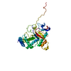
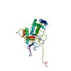
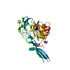
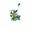
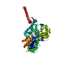
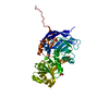
N512P mutant of the DNA binding domain of the Adenovirus 5 ssDNA binding protein
ComponentsE2A DNA-BINDING PROTEIN
KeywordsDNA BINDING PROTEIN / DNA-BINDING PROTEIN / SSDNA BINDING PROTEIN
Function / homology
Function and homology information


viral DNA strand displacement replication / viral DNA genome replication / DNA duplex unwinding / DNA unwinding involved in DNA replication / positive regulation of DNA replication / viral capsid / single-stranded DNA binding / DNA-templated transcription / host cell nucleus / DNA binding ...viral DNA strand displacement replication / viral DNA genome replication / DNA duplex unwinding / DNA unwinding involved in DNA replication / positive regulation of DNA replication / viral capsid / single-stranded DNA binding / DNA-templated transcription / host cell nucleus / DNA binding / zinc ion binding / identical protein binding
Similarity search - Function
Adenovirus Single-stranded Dna-binding Protein, domain 1 / Adenovirus DNA-binding, N-terminal domain / Adenovirus Single-stranded DNA-binding Protein; domain 2 / Adenovirus DNA-binding, C-terminal domain superfamily/Adenovirus DNA-binding, zinc binding domain / Adenovirus DNA-binding, all-alpha domain / Adenovirus DNA-binding, zinc-binding domain / Adenovirus DNA-binding, N-terminal domain superfamily / Adenovirus DNA-binding, C-terminal domain superfamily / Adenovirus DNA-binding, zinc binding domain superfamily / DNA-binding protein, Adenovirus ...Adenovirus Single-stranded Dna-binding Protein, domain 1 / Adenovirus DNA-binding, N-terminal domain / Adenovirus Single-stranded DNA-binding Protein; domain 2 / Adenovirus DNA-binding, C-terminal domain superfamily/Adenovirus DNA-binding, zinc binding domain / Adenovirus DNA-binding, all-alpha domain / Adenovirus DNA-binding, zinc-binding domain / Adenovirus DNA-binding, N-terminal domain superfamily / Adenovirus DNA-binding, C-terminal domain superfamily / Adenovirus DNA-binding, zinc binding domain superfamily / DNA-binding protein, Adenovirus / Viral DNA-binding protein, all alpha domain / Viral DNA-binding protein, zinc binding domain / Alpha-Beta Complex / Orthogonal Bundle / Mainly Alpha / Alpha Beta
Similarity search - Domain/homology
DNA-binding protein / DNA-binding protein
Similarity search - Component
Biological speciesHUMAN ADENOVIRUS 5
MethodX-RAY DIFFRACTION / SYNCHROTRON / MOLECULAR REPLACEMENT / Resolution: 2.3 Å
AuthorsHendle, J. / Kanellopoulos, P.N. / Tucker, P.A.
CitationJournal: To be Published
Title: High Resolution Structures of the Adenovirus Single-Stranded DNA Binding Protein and the N512P Hinge-Region Mutant
Authors: Hendle, J. / Kanellopoulos, P.N. / Tucker, P.A.
History
DepositionFeb 19, 2009Deposition site: PDBE / Processing site: PDBE
Revision 1.0Mar 31, 2010Provider: repository / Type: Initial release
Revision 1.1May 8, 2011Group: Version format compliance
Revision 1.2Jul 13, 2011Group: Version format compliance
Revision 1.3Jul 24, 2019Group: Data collection / Category: diffrn_source / Item: _diffrn_source.pdbx_synchrotron_site
Revision 1.4Dec 13, 2023Group: Data collection / Database references ...Data collection / Database references / Derived calculations / Other / Refinement description
Category: chem_comp_atom / chem_comp_bond ...chem_comp_atom / chem_comp_bond / database_2 / pdbx_database_status / pdbx_initial_refinement_model / pdbx_struct_conn_angle / struct_conn / struct_site
Item: _database_2.pdbx_DOI / _database_2.pdbx_database_accession ..._database_2.pdbx_DOI / _database_2.pdbx_database_accession / _pdbx_database_status.status_code_sf / _pdbx_struct_conn_angle.ptnr1_auth_comp_id / _pdbx_struct_conn_angle.ptnr1_auth_seq_id / _pdbx_struct_conn_angle.ptnr1_label_atom_id / _pdbx_struct_conn_angle.ptnr1_label_comp_id / _pdbx_struct_conn_angle.ptnr1_label_seq_id / _pdbx_struct_conn_angle.ptnr3_auth_comp_id / _pdbx_struct_conn_angle.ptnr3_auth_seq_id / _pdbx_struct_conn_angle.ptnr3_label_atom_id / _pdbx_struct_conn_angle.ptnr3_label_comp_id / _pdbx_struct_conn_angle.ptnr3_label_seq_id / _pdbx_struct_conn_angle.value / _struct_conn.pdbx_dist_value / _struct_conn.ptnr1_auth_comp_id / _struct_conn.ptnr1_auth_seq_id / _struct_conn.ptnr1_label_asym_id / _struct_conn.ptnr1_label_atom_id / _struct_conn.ptnr1_label_comp_id / _struct_conn.ptnr1_label_seq_id / _struct_conn.ptnr2_auth_comp_id / _struct_conn.ptnr2_auth_seq_id / _struct_conn.ptnr2_label_asym_id / _struct_conn.ptnr2_label_atom_id / _struct_conn.ptnr2_label_comp_id / _struct_conn.ptnr2_label_seq_id / _struct_site.pdbx_auth_asym_id / _struct_site.pdbx_auth_comp_id / _struct_site.pdbx_auth_seq_id

-
Structure visualization

Structure viewerMolecule:
MolmilJmol/JSmol

Downloads & links

-
Assembly

Deposited unit
X: E2A DNA-BINDING PROTEIN
hetero molecules


Theoretical massNumber of molelcules
Total (without water)40,0143
Polymers39,8831
Non-polymers1312
Water6,251347
1


  • Idetical with deposited unit
  • defined by author&software
TypeNameSymmetry operationNumber
identity operation1_555x,y,z1
MethodPISA
Unit cell
Length a, b, c (Å)78.320, 75.930, 64.635
Angle α, β, γ (deg.)90.00, 90.00, 90.00
Int Tables number19
Space group name H-MP212121

-
Components

#1: Protein E2A DNA-BINDING PROTEIN / ADENOVIRUS 5 SSDNA BINDING PROTEIN


Mass: 39882.758 Da / Num. of mol.: 1 / Fragment: C-TERMINAL DNA BINDING DOMAIN, RESIDUES 174-529 / Mutation: YES
Source method: isolated from a genetically manipulated source
Source: (gene. exp.) HUMAN ADENOVIRUS 5 / Plasmid: PETM11 / Production host: ESCHERICHIA COLI (E. coli) / Strain (production host): BL21 / References: UniProt: Q2KS06, UniProt: P03265*PLUS
#2: Chemical ChemComp-ZN / ZINC ION


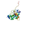
Mass: 65.409 Da / Num. of mol.: 2 / Source method: obtained synthetically / Formula: Zn
#3: Water ChemComp-HOH / water


Mass: 18.015 Da / Num. of mol.: 347 / Source method: isolated from a natural source / Formula: H2O
Compound detailsENGINEERED RESIDUE IN CHAIN X, ASN 512 TO PRO
Sequence detailsN512P MUTATION

-
Experimental details

-
Experiment

ExperimentMethod: X-RAY DIFFRACTION / Number of used crystals: 1

-
Sample preparation

CrystalDensity Matthews: 2.4 Å3/Da / Density % sol: 48.8 % / Description: NONE
Crystal growDetails: 2.0M NANO3 WITH SODIUM ACETATE BUFFER AT PH5.8

-
Data collection

DiffractionMean temperature: 100 K
Diffraction sourceSource: SYNCHROTRON / Site: EMBL/DESY, HAMBURG / Beamline: BW7B / Wavelength: 0.838
DetectorType: MARRESEARCH / Detector: IMAGE PLATE / Details: MIRRORS
RadiationMonochromator: SI(111) / Protocol: SINGLE WAVELENGTH / Monochromatic (M) / Laue (L): M / Scattering type: x-ray
Radiation wavelengthWavelength: 0.838 Å / Relative weight: 1
ReflectionResolution: 2.3→30 Å / Num. obs: 16993 / % possible obs: 88.6 % / Observed criterion σ(I): -3 / Redundancy: 4.5 % / Rmerge(I) obs: 0.08 / Net I/σ(I): 17.6
Reflection shellResolution: 2.3→2.45 Å / Redundancy: 4.6 % / Rmerge(I) obs: 0.11 / Mean I/σ(I) obs: 8 / % possible all: 97.4

-
Processing

Software
NameVersionClassification
REFMAC5.5.0072refinement
DENZOdata reduction
SCALEPACKdata scaling
REFMACphasing
RefinementMethod to determine structure: MOLECULAR REPLACEMENT
Starting model: PDB ENTRY 1ADT

1adt
PDB Unreleased entry


Resolution: 2.3→29.87 Å / Cor.coef. Fo:Fc: 0.951 / Cor.coef. Fo:Fc free: 0.911 / SU B: 5.796 / SU ML: 0.145 / Cross valid method: THROUGHOUT / ESU R: 0.305 / ESU R Free: 0.244 / Stereochemistry target values: MAXIMUM LIKELIHOOD / Details: HYDROGENS HAVE BEEN ADDED IN THE RIDING POSITIONS.
RfactorNum. reflection% reflectionSelection details
Rfree0.246 877 5.2 %RANDOM
Rwork0.167 ---
obs0.171 16116 96.1 %-
Solvent computationIon probe radii: 0.8 Å / Shrinkage radii: 0.8 Å / VDW probe radii: 1.4 Å / Solvent model: MASK
Displacement parametersBiso mean: 31.61 Å2
Baniso -1Baniso -2Baniso -3
1-0.02 Å2-0 Å20 Å2
2---0.03 Å2-0 Å2
3---0.01 Å2
Refinement stepCycle: LAST / Resolution: 2.3→29.87 Å
ProteinNucleic acidLigandSolventTotal
Num. atoms2477 0 2 347 2826
Refine LS restraints
Refine-IDTypeDev idealDev ideal targetNumber
X-RAY DIFFRACTIONr_bond_refined_d0.0220.0222556
X-RAY DIFFRACTIONr_bond_other_d
X-RAY DIFFRACTIONr_angle_refined_deg1.9341.9443462
X-RAY DIFFRACTIONr_angle_other_deg
X-RAY DIFFRACTIONr_dihedral_angle_1_deg6.6935314
X-RAY DIFFRACTIONr_dihedral_angle_2_deg36.38624.31116
X-RAY DIFFRACTIONr_dihedral_angle_3_deg19.46315424
X-RAY DIFFRACTIONr_dihedral_angle_4_deg19.5461511
X-RAY DIFFRACTIONr_chiral_restr0.1570.2375
X-RAY DIFFRACTIONr_gen_planes_refined0.0090.0211945
X-RAY DIFFRACTIONr_gen_planes_other
X-RAY DIFFRACTIONr_nbd_refined
X-RAY DIFFRACTIONr_nbd_other
X-RAY DIFFRACTIONr_nbtor_refined
X-RAY DIFFRACTIONr_nbtor_other
X-RAY DIFFRACTIONr_xyhbond_nbd_refined
X-RAY DIFFRACTIONr_xyhbond_nbd_other
X-RAY DIFFRACTIONr_metal_ion_refined
X-RAY DIFFRACTIONr_metal_ion_other
X-RAY DIFFRACTIONr_symmetry_vdw_refined
X-RAY DIFFRACTIONr_symmetry_vdw_other
X-RAY DIFFRACTIONr_symmetry_hbond_refined
X-RAY DIFFRACTIONr_symmetry_hbond_other
X-RAY DIFFRACTIONr_symmetry_metal_ion_refined
X-RAY DIFFRACTIONr_symmetry_metal_ion_other
X-RAY DIFFRACTIONr_mcbond_it1.1161.51584
X-RAY DIFFRACTIONr_mcbond_other
X-RAY DIFFRACTIONr_mcangle_it2.08922547
X-RAY DIFFRACTIONr_mcangle_other
X-RAY DIFFRACTIONr_scbond_it3.1593972
X-RAY DIFFRACTIONr_scbond_other
X-RAY DIFFRACTIONr_scangle_it5.0114.5915
X-RAY DIFFRACTIONr_scangle_other
X-RAY DIFFRACTIONr_long_range_B_refined
X-RAY DIFFRACTIONr_long_range_B_other
X-RAY DIFFRACTIONr_rigid_bond_restr
X-RAY DIFFRACTIONr_sphericity_free
X-RAY DIFFRACTIONr_sphericity_bonded
LS refinement shellResolution: 2.3→2.36 Å / Total num. of bins used: 20 /
RfactorNum. reflection
Rfree0.313 67
Rwork0.185 1142

+
About Yorodumi

-
News

-
Feb 9, 2022. New format data for meta-information of EMDB entries

New format data for meta-information of EMDB entries

  • Version 3 of the EMDB header file is now the official format.
  • The previous official version 1.9 will be removed from the archive.

Related info.:EMDB header

External links:wwPDB to switch to version 3 of the EMDB data model

-
Aug 12, 2020. Covid-19 info

Covid-19 info

URL: https://pdbjlvh1.pdbj.org/emnavi/covid19.php

New page: Covid-19 featured information page in EM Navigator.

Related info.:Covid-19 info / Mar 5, 2020. Novel coronavirus structure data

+
Mar 5, 2020. Novel coronavirus structure data

Novel coronavirus structure data

Related info.:Yorodumi Speices / Aug 12, 2020. Covid-19 info

External links:COVID-19 featured content - PDBj / Molecule of the Month (242):Coronavirus Proteases

+
Jan 31, 2019. EMDB accession codes are about to change! (news from PDBe EMDB page)

EMDB accession codes are about to change! (news from PDBe EMDB page)

  • The allocation of 4 digits for EMDB accession codes will soon come to an end. Whilst these codes will remain in use, new EMDB accession codes will include an additional digit and will expand incrementally as the available range of codes is exhausted. The current 4-digit format prefixed with “EMD-” (i.e. EMD-XXXX) will advance to a 5-digit format (i.e. EMD-XXXXX), and so on. It is currently estimated that the 4-digit codes will be depleted around Spring 2019, at which point the 5-digit format will come into force.
  • The EM Navigator/Yorodumi systems omit the EMD- prefix.

Related info.:Q: What is EMD? / ID/Accession-code notation in Yorodumi/EM Navigator

External links:EMDB Accession Codes are Changing Soon! / Contact to PDBj

+
Jul 12, 2017. Major update of PDB

Major update of PDB

  • wwPDB released updated PDB data conforming to the new PDBx/mmCIF dictionary.
  • This is a major update changing the version number from 4 to 5, and with Remediation, in which all the entries are updated.
  • In this update, many items about electron microscopy experimental information are reorganized (e.g. em_software).
  • Now, EM Navigator and Yorodumi are based on the updated data.

External links:wwPDB Remediation / Enriched Model Files Conforming to OneDep Data Standards Now Available in the PDB FTP Archive

-
Yorodumi

Thousand views of thousand structures

  • Yorodumi is a browser for structure data from EMDB, PDB, SASBDB, etc.
  • This page is also the successor to EM Navigator detail page, and also detail information page/front-end page for Omokage search.
  • The word "yorodu" (or yorozu) is an old Japanese word meaning "ten thousand". "mi" (miru) is to see.

Related info.:EMDB / PDB / SASBDB / Comparison of 3 databanks / Yorodumi Search / Aug 31, 2016. New EM Navigator & Yorodumi / Yorodumi Papers / Jmol/JSmol / Function and homology information / Changes in new EM Navigator and Yorodumi

Read more