 Movie
Movie Controller


+ Open data

- Basic information
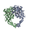
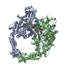
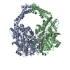
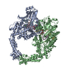
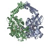
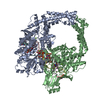
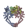
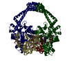
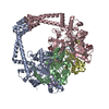
| Entry | Database: PDB / ID: 4g0v | ||||||
|---|---|---|---|---|---|---|---|
| Title | Human topoisomerase iibeta in complex with DNA and mitoxantrone | ||||||
 Components Components |
| ||||||
 Keywords Keywords | ISOMERASE/DNA/ISOMERASE INHIBITOR / TOPRIM domain / winged-helix domain / coiled-coil domain / DNA-binding and cleavage / nucleus / ISOMERASE-DNA-ISOMERASE INHIBITOR complex | ||||||
| Function / homology |  Function and homology information Function and homology informationpositive regulation of single stranded viral RNA replication via double stranded DNA intermediate / sister chromatid segregation / resolution of meiotic recombination intermediates / DNA topoisomerase type II (double strand cut, ATP-hydrolyzing) activity / DNA topoisomerase (ATP-hydrolysing) / positive regulation of double-strand break repair via nonhomologous end joining / cellular response to ATP / forebrain development / DNA topological change / SUMOylation of DNA replication proteins ...positive regulation of single stranded viral RNA replication via double stranded DNA intermediate / sister chromatid segregation / resolution of meiotic recombination intermediates / DNA topoisomerase type II (double strand cut, ATP-hydrolyzing) activity / DNA topoisomerase (ATP-hydrolysing) / positive regulation of double-strand break repair via nonhomologous end joining / cellular response to ATP / forebrain development / DNA topological change / SUMOylation of DNA replication proteins / ribonucleoprotein complex binding / axonogenesis / B cell differentiation / neuron migration / cellular response to hydrogen peroxide / cellular senescence / ribonucleoprotein complex / chromatin binding / nucleolus / DNA binding / nucleoplasm / ATP binding / metal ion binding / nucleus / cytosol Similarity search - Function | ||||||
| Biological species |  Homo sapiens (human) Homo sapiens (human) | ||||||
| Method |  X-RAY DIFFRACTION / SYNCHROTRON / MOLECULAR REPLACEMENT / Resolution: 2.548 Å X-RAY DIFFRACTION / SYNCHROTRON / MOLECULAR REPLACEMENT / Resolution: 2.548 Å | ||||||
 Authors Authors | Wu, C.C. / Li, T.K. / Li, Y.C. / Chan, N.L. | ||||||
 Citation Citation | Journal: Nucleic Acids Res. / Year: 2013 Title: On the structural basis and design guidelines for type II topoisomerase-targeting anticancer drugs Authors: Wu, C.C. / Li, Y.C. / Wang, Y.R. / Li, T.K. / Chan, N.L. | ||||||
| History |
|
- Structure visualization
| Structure viewer | Molecule: MolmilJmol/JSmol |
|---|
- Downloads & links
-Download
| PDBx/mmCIF format | 4g0v.cif.gz | 327.6 KB | Display | PDBx/mmCIF format |
|---|---|---|---|---|
| PDB format | pdb4g0v.ent.gz | 252.1 KB | Display | PDB format |
| PDBx/mmJSON format | 4g0v.json.gz | Tree view | PDBx/mmJSON format | |
| Others |  Other downloads Other downloads |
-Validation report
| Arichive directory | https://data.pdbj.org/pub/pdb/validation_reports/g0/4g0vftp://data.pdbj.org/pub/pdb/validation_reports/g0/4g0v | HTTPS FTP |
|---|
-Related structure data
| Related structure data |  4g0uC  4g0wC  4j3nC  3qx3S S: Starting model for refinement C: citing same article ( |
|---|---|
| Similar structure data |

-Links
 PDBj
PDBj


- Assembly
| Deposited unit | 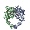
| ||||||||
|---|---|---|---|---|---|---|---|---|---|
| 1 |
| ||||||||
| Unit cell |
|
-Components
-Protein , 1 types, 2 molecules AB
| #1: Protein | Mass: 91928.367 Da / Num. of mol.: 2 / Fragment: HTOP2BETA CLEAVAGE CORE, UNP RESIDUES 450-1206 Source method: isolated from a genetically manipulated source Source: (gene. exp.) Homo sapiens (human) / Gene: TOP2B / Plasmid: PET51B / Production host:  |
|---|
-DNA chain , 2 types, 4 molecules CEDF
| #2: DNA chain | Mass: 2436.619 Da / Num. of mol.: 2 / Source method: obtained synthetically #3: DNA chain | Mass: 3654.378 Da / Num. of mol.: 2 / Source method: obtained synthetically |
|---|
-Non-polymers , 3 types, 567 molecules 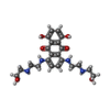


| #4: Chemical |  Mass: 444.481 Da / Num. of mol.: 2 / Source method: obtained synthetically / Formula: C22H28N4O6 / Comment: antineoplastic*YM Mass: 444.481 Da / Num. of mol.: 2 / Source method: obtained synthetically / Formula: C22H28N4O6 / Comment: antineoplastic*YM#5: Chemical | ChemComp-MG /  Mass: 24.305 Da / Num. of mol.: 6 / Source method: obtained synthetically / Formula: Mg Mass: 24.305 Da / Num. of mol.: 6 / Source method: obtained synthetically / Formula: Mg#6: Water | ChemComp-HOH / | Mass: 18.015 Da / Num. of mol.: 559 / Source method: isolated from a natural source / Formula: H2O |
|---|
-Details
| Has protein modification | Y |
|---|---|
| Sequence details | THE SEQUENCE OF DNA USED IN THE EXPERIMENT WAS 5'-AGCCGAGCTGCAGCTCGGCT-3'. AND THE DNAS WERE ...THE SEQUENCE OF DNA USED IN THE EXPERIMENT |
-Experimental details
-Experiment
| Experiment | Method: X-RAY DIFFRACTION / Number of used crystals: 1 |
|---|
- Sample preparation
| Crystal | Density Matthews: 3.16 Å3/Da / Density % sol: 61.13 % |
|---|---|
| Crystal grow | Temperature: 277 K / Method: vapor diffusion, hanging drop / pH: 5.8 Details: 100mM magnesium acetate, 50mM 2-(N-morpholino)ethanesulfonic acid pH 5.8, 22% 2-methyl-2,4-pentanediol (MPD), VAPOR DIFFUSION, HANGING DROP, temperature 277K |
-Data collection
| Diffraction | Mean temperature: 100 K |
|---|---|
| Diffraction source | Source: SYNCHROTRON / Site: NSRRC  / Beamline: BL13C1 / Wavelength: 0.97622 Å / Beamline: BL13C1 / Wavelength: 0.97622 Å |
| Detector | Type: RAYONIX MX300HE / Detector: CCD / Date: Dec 25, 2010 |
| Radiation | Monochromator: HORIZONTALLY FOCUSING SINGLE CRYSTAL MONOCHROMATOR (CRYSTAL TYPE SI(1 1 1)) Protocol: SINGLE WAVELENGTH / Monochromatic (M) / Laue (L): M / Scattering type: x-ray |
| Radiation wavelength | Wavelength: 0.97622 Å / Relative weight: 1 |
| Reflection | Resolution: 2.548→30 Å / Num. all: 79306 / Num. obs: 78830 / % possible obs: 99.4 % / Observed criterion σ(F): 1 / Observed criterion σ(I): 1 / Redundancy: 3.6 % / Biso Wilson estimate: 42.58 Å2 / Rsym value: 0.07 / Net I/σ(I): 12.1 |
| Reflection shell | Resolution: 2.55→2.59 Å / Redundancy: 3.5 % / Rmerge(I) obs: 0.49 / Mean I/σ(I) obs: 2.5 / Num. unique all: 3892 / Rsym value: 0.49 / % possible all: 97.3 |
- Processing
| Software |
| |||||||||||||||||||||||||||||||||||||||||||||||||||||||||||||||||||||||||||||
|---|---|---|---|---|---|---|---|---|---|---|---|---|---|---|---|---|---|---|---|---|---|---|---|---|---|---|---|---|---|---|---|---|---|---|---|---|---|---|---|---|---|---|---|---|---|---|---|---|---|---|---|---|---|---|---|---|---|---|---|---|---|---|---|---|---|---|---|---|---|---|---|---|---|---|---|---|---|---|
| Refinement | Method to determine structure: MOLECULAR REPLACEMENT Starting model: PDB ENTRY 3QX3 Resolution: 2.548→27.941 Å / Occupancy max: 1 / Occupancy min: 0.29 / FOM work R set: 0.8556 / SU ML: 0.28 / σ(F): 1.35 / Phase error: 21.86 / Stereochemistry target values: ML
| |||||||||||||||||||||||||||||||||||||||||||||||||||||||||||||||||||||||||||||
| Solvent computation | Shrinkage radii: 0.7 Å / VDW probe radii: 1 Å / Solvent model: FLAT BULK SOLVENT MODEL | |||||||||||||||||||||||||||||||||||||||||||||||||||||||||||||||||||||||||||||
| Displacement parameters | Biso max: 131.86 Å2 / Biso mean: 33.3113 Å2 / Biso min: 8.43 Å2 | |||||||||||||||||||||||||||||||||||||||||||||||||||||||||||||||||||||||||||||
| Refinement step | Cycle: LAST / Resolution: 2.548→27.941 Å
| |||||||||||||||||||||||||||||||||||||||||||||||||||||||||||||||||||||||||||||
| Refine LS restraints |
| |||||||||||||||||||||||||||||||||||||||||||||||||||||||||||||||||||||||||||||
| LS refinement shell | Refine-ID: X-RAY DIFFRACTION / Total num. of bins used: 10
|
