 ムービー
ムービー コントローラー


+ データを開く

- 基本情報
基本情報
| 登録情報 | データベース: PDB / ID: 1gr5 | ||||||
|---|---|---|---|---|---|---|---|
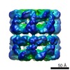
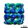
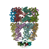
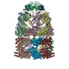
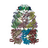
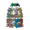
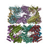
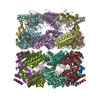
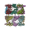
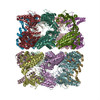
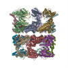
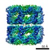
| タイトル | Solution Structure of apo GroEL by Cryo-Electron microscopy | ||||||
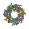
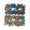
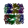
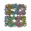
 要素 要素 | 60 KDA CHAPERONIN | ||||||
 キーワード キーワード | CHAPERONE | ||||||
| 機能・相同性 |  機能・相同性情報 機能・相同性情報GroEL-GroES complex / chaperonin ATPase / cell cycle / virion assembly / chaperone cofactor-dependent protein refolding / isomerase activity / ATP-dependent protein folding chaperone / response to radiation / unfolded protein binding / protein folding ...GroEL-GroES complex / chaperonin ATPase / cell cycle / virion assembly / chaperone cofactor-dependent protein refolding / isomerase activity / ATP-dependent protein folding chaperone / response to radiation / unfolded protein binding / protein folding / protein refolding / response to heat / cell division / magnesium ion binding / ATP hydrolysis activity / ATP binding / identical protein binding / membrane / cytoplasm / cytosol 類似検索 - 分子機能 | ||||||
| 生物種 |  | ||||||
| 手法 | 電子顕微鏡法 / 単粒子再構成法 / クライオ電子顕微鏡法 / 解像度: 7.9 Å | ||||||
 データ登録者 データ登録者 | Ranson, N.A. / Farr, G.W. / Roseman, A.M. / Gowen, B. / Fenton, W.A. / Horwich, A.L. / Saibil, H.R. | ||||||
 引用 引用 | ジャーナル: Cell / 年: 2001 タイトル: ATP-bound states of GroEL captured by cryo-electron microscopy. 著者: N A Ranson / G W Farr / A M Roseman / B Gowen / W A Fenton / A L Horwich / H R Saibil /  要旨: The chaperonin GroEL drives its protein-folding cycle by cooperatively binding ATP to one of its two rings, priming that ring to become folding-active upon GroES binding, while simultaneously ...The chaperonin GroEL drives its protein-folding cycle by cooperatively binding ATP to one of its two rings, priming that ring to become folding-active upon GroES binding, while simultaneously discharging the previous folding chamber from the opposite ring. The GroEL-ATP structure, determined by cryo-EM and atomic structure fitting, shows that the intermediate domains rotate downward, switching their intersubunit salt bridge contacts from substrate binding to ATP binding domains. These observations, together with the effects of ATP binding to a GroEL-GroES-ADP complex, suggest structural models for the ATP-induced reduction in affinity for polypeptide and for cooperativity. The model for cooperativity, based on switching of intersubunit salt bridge interactions around the GroEL ring, may provide general insight into cooperativity in other ring complexes and molecular machines. #1: ジャーナル: Nature / 年: 1997タイトル: The crystal structure of the asymmetric GroEL-GroES-(ADP)7 chaperonin complex. 著者: Z Xu / A L Horwich / P B Sigler /  要旨: Chaperonins assist protein folding with the consumption of ATP. They exist as multi-subunit protein assemblies comprising rings of subunits stacked back to back. In Escherichia coli, asymmetric ...Chaperonins assist protein folding with the consumption of ATP. They exist as multi-subunit protein assemblies comprising rings of subunits stacked back to back. In Escherichia coli, asymmetric intermediates of GroEL are formed with the co-chaperonin GroES and nucleotides bound only to one of the seven-subunit rings (the cis ring) and not to the opposing ring (the trans ring). The structure of the GroEL-GroES-(ADP)7 complex reveals how large en bloc movements of the cis ring's intermediate and apical domains enable bound GroES to stabilize a folding chamber with ADP confined to the cis ring. Elevation and twist of the apical domains double the volume of the central cavity and bury hydrophobic peptide-binding residues in the interface with GroES, as well as between GroEL subunits, leaving a hydrophilic cavity lining that is conducive to protein folding. An inward tilt of the cis equatorial domain causes an outward tilt in the trans ring that opposes the binding of a second GroES. When combined with new functional results, this negative allosteric mechanism suggests a model for an ATP-driven folding cycle that requires a double toroid. #2: ジャーナル: Nat Struct Biol / 年: 1996 タイトル: The 2.4 A crystal structure of the bacterial chaperonin GroEL complexed with ATP gamma S. 著者: D C Boisvert / J Wang / Z Otwinowski / A L Horwich / P B Sigler / 要旨: GroEL is a bacterial chaperonin of 14 identical subunits required to help fold newly synthesized proteins. The crystal structure of GroEL with ATP gamma S bound to each subunit shows that ATP binds ...GroEL is a bacterial chaperonin of 14 identical subunits required to help fold newly synthesized proteins. The crystal structure of GroEL with ATP gamma S bound to each subunit shows that ATP binds to a novel pocket, whose primary sequence is highly conserved among chaperonins. Interaction of Mg2+ and ATP involves phosphate oxygens of the alpha-, beta- and gamma-phosphates, which is unique for known structures of nucleotide-binding proteins. Although bound ATP induces modest conformational shifts in the equatorial domain, the stereochemistry that functionally coordinates GroEL's affinity for nucleotides, polypeptide, and GroES remains uncertain. #3: ジャーナル: Nature / 年: 1994タイトル: The crystal structure of the bacterial chaperonin GroEL at 2.8 A. 著者: K Braig / Z Otwinowski / R Hegde / D C Boisvert / A Joachimiak / A L Horwich / P B Sigler / 要旨: The crystal structure of Escherichia coli GroEL shows a porous cylinder of 14 subunits made of two nearly 7-fold rotationally symmetrical rings stacked back-to-back with dyad symmetry. The subunits ...The crystal structure of Escherichia coli GroEL shows a porous cylinder of 14 subunits made of two nearly 7-fold rotationally symmetrical rings stacked back-to-back with dyad symmetry. The subunits consist of three domains: a large equatorial domain that forms the foundation of the assembly at its waist and holds the rings together; a large loosely structured apical domain that forms the ends of the cylinder; and a small slender intermediate domain that connects the two, creating side windows. The three-dimensional structure places most of the mutationally defined functional sites on the channel walls and its outward invaginations, and at the ends of the cylinder. | ||||||
| 履歴 |
| ||||||
| Remark 700 | SHEET THE SHEET STRUCTURE OF THIS MOLECULE IS BIFURCATED. IN ORDER TO REPRESENT THIS FEATURE IN ... SHEET THE SHEET STRUCTURE OF THIS MOLECULE IS BIFURCATED. IN ORDER TO REPRESENT THIS FEATURE IN THE SHEET RECORDS BELOW, TWO SHEETS ARE DEFINED. |
- 構造の表示
構造の表示
| ムービー |
ムービービューア |
|---|---|
| 構造ビューア | 分子: MolmilJmol/JSmol |
- ダウンロードとリンク
-ダウンロード
| PDBx/mmCIF形式 | 1gr5.cif.gz | 1.2 MB | 表示 | PDBx/mmCIF形式 |
|---|---|---|---|---|
| PDB形式 | pdb1gr5.ent.gz | 1 MB | 表示 | PDB形式 |
| PDBx/mmJSON形式 | 1gr5.json.gz | ツリー表示 | PDBx/mmJSON形式 | |
| その他 |  その他のダウンロード その他のダウンロード |
-検証レポート
| 文書・要旨 | 1gr5_validation.pdf.gz | 1.1 MB | 表示 | wwPDB検証レポート |
|---|---|---|---|---|
| 文書・詳細版 | 1gr5_full_validation.pdf.gz | 1.3 MB | 表示 | |
| XML形式データ | 1gr5_validation.xml.gz | 211.1 KB | 表示 | |
| CIF形式データ | 1gr5_validation.cif.gz | 304.6 KB | 表示 | |
| アーカイブディレクトリ | https://data.pdbj.org/pub/pdb/validation_reports/gr/1gr5ftp://data.pdbj.org/pub/pdb/validation_reports/gr/1gr5 | HTTPS FTP |
-関連構造データ


-リンク
 PDBj
PDBj
- 集合体
集合体
| 登録構造単位 | 
|
|---|---|
| 1 |
|
-要素
| #1: タンパク質 | 分子量: 57188.410 Da / 分子数: 14 / 変異: YES / 由来タイプ: 組換発現 / 由来: (組換発現) 構成要素の詳細 | GROEL IS A HOMOOLIGOMER OF FOURTEEN SUBUNITS ARRANGED IN A DOUBLE RING STRUCTURE. ENGINEERED ...GROEL IS A HOMOOLIGOM | 配列の詳細 | MET 1 IN ALL CHAINS HAS BEEN POST-TRANSLATIO | |
|---|
-実験情報
-実験
| 実験 | 手法: 電子顕微鏡法 |
|---|---|
| EM実験 | 試料の集合状態: PARTICLE / 3次元再構成法: 単粒子再構成法 |
- 試料調製
試料調製
| 構成要素 | 名称: MOLECULAR CHAPERONE / タイプ: COMPLEX |
|---|---|
| 緩衝液 | 名称: HEPES / pH: 7.5 / 詳細: HEPES |
| 試料 | 濃度: 0.7 mg/ml / 包埋: NO / シャドウイング: NO / 染色: NO / 凍結: YES |
| 試料支持 | 詳細: HOLEY CARBON |
| 急速凍結 | 装置: HOMEMADE PLUNGER / 凍結剤: ETHANE / 詳細: LIQUID ETHANE |
| 結晶化 | *PLUS 手法: cryo-electron microscopy |
- 電子顕微鏡撮影
| 顕微鏡 | モデル: FEI/PHILIPS CM200T / 日付: 1997年2月1日 |
|---|---|
| 電子銃 | 電子線源:  FIELD EMISSION GUN / 加速電圧: 200 kV / 照射モード: FLOOD BEAM FIELD EMISSION GUN / 加速電圧: 200 kV / 照射モード: FLOOD BEAM |
| 電子レンズ | モード: BRIGHT FIELD / 倍率(公称値): 38000 X / 倍率(補正後): 40200 X / 最大 デフォーカス(公称値): 1900 nm / 最小 デフォーカス(公称値): 800 nm |
| 試料ホルダ | 温度: 95 K / 傾斜角・最大: 0 ° / 傾斜角・最小: 0 ° |
| 撮影 | 電子線照射量: 20 e/Å2 / フィルム・検出器のモデル: KODAK SO-163 FILM |
| 画像スキャン | デジタル画像の数: 50 |
| 放射波長 | 相対比: 1 |
- 解析
解析
| EMソフトウェア |
| ||||||||||||
|---|---|---|---|---|---|---|---|---|---|---|---|---|---|
| CTF補正 | 詳細: CLASS AVERAGES | ||||||||||||
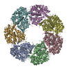
| 対称性 | 点対称性: D7 (2回x7回 2面回転対称) | ||||||||||||
| 3次元再構成 | 手法: MODEL-BASED ANGULAR REFINEMENT / 解像度: 7.9 Å / 解像度の算出法: FSC 0.5 CUT-OFF / 粒子像の数: 8728 / ピクセルサイズ(公称値): 1.82 Å / ピクセルサイズ(実測値): 1.74 Å 詳細: THE THREE DOMAINS FROM EACH SUBUNIT (PDB 1DER) WERE FITTED SEPARATELY INTO EM DENSITY OF WILD TYPE UNLIGANDED GROEL. THE MUTATIONS LISTED ARE PRESENT IN THE FITTED COORDINATES AND NOT IN THE ...詳細: THE THREE DOMAINS FROM EACH SUBUNIT (PDB 1DER) WERE FITTED SEPARATELY INTO EM DENSITY OF WILD TYPE UNLIGANDED GROEL. THE MUTATIONS LISTED ARE PRESENT IN THE FITTED COORDINATES AND NOT IN THE MOLECULE WHICH GENERATED THE EM MAP. THE QUOTED RESOLUTION IS AT 3SIGMA - THE RESOLUTION AT FSC=0.5 IS 10.8A THIS ENTRY REPRESENTS THE COMPLETE BIOMOLECULE. 対称性のタイプ: POINT | ||||||||||||
| 原子モデル構築 | プロトコル: OTHER / 空間: REAL 詳細: METHOD--LOCAL CORRELATION REFINEMENT PROTOCOL--X-RAY | ||||||||||||
| 原子モデル構築 | PDB-ID: 1DER 1der Accession code: 1DER / Source name: PDB / タイプ: experimental model | ||||||||||||
| 精密化 | 最高解像度: 7.9 Å | ||||||||||||
| 精密化ステップ | サイクル: LAST / 最高解像度: 7.9 Å
| ||||||||||||
| 精密化 | *PLUS 最高解像度: 7.9 Å | ||||||||||||
| 溶媒の処理 | *PLUS | ||||||||||||
| 原子変位パラメータ | *PLUS |
