 Movie
Movie Controller

[English] 日本語
 Yorodumi
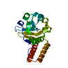
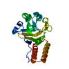
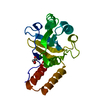
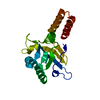
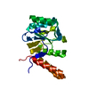
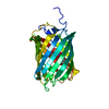
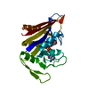
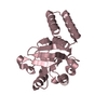
- PDB-1kid: GROEL (HSP60 CLASS) FRAGMENT (APICAL DOMAIN) COMPRISING RESIDUES ... -
+ Open data

- Basic information
| Entry | Database: PDB / ID: 1kid | ||||||
|---|---|---|---|---|---|---|---|
| Title | GROEL (HSP60 CLASS) FRAGMENT (APICAL DOMAIN) COMPRISING RESIDUES 191-376, MUTANT WITH ALA 262 REPLACED WITH LEU AND ILE 267 REPLACED WITH MET | ||||||
 Components Components | GROEL (HSP60 CLASS) | ||||||
 Keywords Keywords | CHAPERONE / HSP60 / GROEL / CELL DIVISION / ATP-BINDING / PHOSPHORYLATION | ||||||
| Function / homology |  Function and homology information Function and homology informationGroEL-GroES complex / chaperonin ATPase / virion assembly / isomerase activity / ATP-dependent protein folding chaperone / response to radiation / : / protein refolding / response to heat / protein folding ...GroEL-GroES complex / chaperonin ATPase / virion assembly / isomerase activity / ATP-dependent protein folding chaperone / response to radiation / : / protein refolding / response to heat / protein folding / magnesium ion binding / ATP hydrolysis activity / ATP binding / membrane / identical protein binding / cytosol Similarity search - Function | ||||||
| Biological species |  | ||||||
| Method |  X-RAY DIFFRACTION / SYNCHROTRON / MOLECULAR REPLACEMENT / Resolution: 1.7 Å X-RAY DIFFRACTION / SYNCHROTRON / MOLECULAR REPLACEMENT / Resolution: 1.7 Å | ||||||
 Authors Authors | Buckle, A.M. / Fersht, A.R. | ||||||
 Citation Citation | Journal: Proc.Natl.Acad.Sci.USA / Year: 1997 Title: A structural model for GroEL-polypeptide recognition. Authors: Buckle, A.M. / Zahn, R. / Fersht, A.R. | ||||||
| History |
|
- Structure visualization
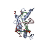
| Structure viewer | Molecule: MolmilJmol/JSmol |
|---|
- Downloads & links
-Download
| PDBx/mmCIF format | 1kid.cif.gz | 56.2 KB | Display | PDBx/mmCIF format |
|---|---|---|---|---|
| PDB format | pdb1kid.ent.gz | 39.9 KB | Display | PDB format |
| PDBx/mmJSON format | 1kid.json.gz | Tree view | PDBx/mmJSON format | |
| Others |  Other downloads Other downloads |
-Validation report
| Arichive directory | https://data.pdbj.org/pub/pdb/validation_reports/ki/1kidftp://data.pdbj.org/pub/pdb/validation_reports/ki/1kid | HTTPS FTP |
|---|
-Related structure data
| Related structure data | 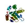 1jonS S: Starting model for refinement |
|---|---|
| Similar structure data |

-Links
 PDBj
PDBj
- Assembly
| Deposited unit | 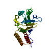
| ||||||||
|---|---|---|---|---|---|---|---|---|---|
| 1 |
| ||||||||
| Unit cell |
| ||||||||
| Details | RESIDUES 184 - 190 (PART OF A FUSED N-TERMINAL TAG) BINDS IN THE PUTATIVE POLYPEPTIDE-BINDING SITE OF A NEIGHBORING MOLECULE IN THE CRYSTAL LATTICE. THIS "MINI-CHAPERONE - PEPTIDE COMPLEX" CAN BE GENERATED BY APPLYING THE TRANSFORMATION 1/2 - X, -Y, 1/2+Z TO THE N-TERMINAL TAG (RESIDUES 184 - 190) AND COMBINING THE RESULTING COORDINATES WITH RESIDUES. |
-Components
| #1: Protein | Mass: 22060.316 Da / Num. of mol.: 1 / Fragment: APICAL DOMAIN, RESIDUES 191 - 376 / Mutation: A262L, I267M Source method: isolated from a genetically manipulated source Source: (gene. exp.) |
|---|---|
| #2: Water | ChemComp-HOH /  Mass: 18.015 Da / Num. of mol.: 292 / Source method: isolated from a natural source / Formula: H2O Mass: 18.015 Da / Num. of mol.: 292 / Source method: isolated from a natural source / Formula: H2O |
-Experimental details
-Experiment
| Experiment | Method: X-RAY DIFFRACTION / Number of used crystals: 1 |
|---|
- Sample preparation
| Crystal | Density Matthews: 2.6 Å3/Da / Density % sol: 52.52 % | ||||||||||||||||||||||||||||||||||||||||||||||||
|---|---|---|---|---|---|---|---|---|---|---|---|---|---|---|---|---|---|---|---|---|---|---|---|---|---|---|---|---|---|---|---|---|---|---|---|---|---|---|---|---|---|---|---|---|---|---|---|---|---|
| Crystal grow | pH: 8.5 / Details: pH 8.5 | ||||||||||||||||||||||||||||||||||||||||||||||||
| Crystal grow | *PLUS Method: vapor diffusion, hanging drop | ||||||||||||||||||||||||||||||||||||||||||||||||
| Components of the solutions | *PLUS
|
-Data collection
| Diffraction | Mean temperature: 100 K |
|---|---|
| Diffraction source | Source: SYNCHROTRON / Site: EMBL/DESY, HAMBURG  / Beamline: X31 / Wavelength: 1.07 / Beamline: X31 / Wavelength: 1.07 |
| Detector | Type: MARRESEARCH / Detector: IMAGE PLATE / Date: Jun 16, 1996 |
| Radiation | Monochromatic (M) / Laue (L): M / Scattering type: x-ray |
| Radiation wavelength | Wavelength: 1.07 Å / Relative weight: 1 |
| Reflection | Resolution: 1.7→12.8 Å / Num. obs: 25290 / % possible obs: 97.6 % / Observed criterion σ(I): 3 / Redundancy: 4.9 % / Biso Wilson estimate: 16.5 Å2 / Rmerge(I) obs: 0.047 / Net I/σ(I): 11.8 |
| Reflection shell | Resolution: 1.7→1.8 Å / Redundancy: 4.4 % / Rmerge(I) obs: 0.217 / Mean I/σ(I) obs: 3.2 / % possible all: 85.6 |
| Reflection | *PLUS Num. measured all: 260633 |
| Reflection shell | *PLUS % possible obs: 85.6 % |
- Processing
| Software |
| ||||||||||||||||||||||||||||||||||||||||||||||||||||||||||||||||||||||||||||||||||||
|---|---|---|---|---|---|---|---|---|---|---|---|---|---|---|---|---|---|---|---|---|---|---|---|---|---|---|---|---|---|---|---|---|---|---|---|---|---|---|---|---|---|---|---|---|---|---|---|---|---|---|---|---|---|---|---|---|---|---|---|---|---|---|---|---|---|---|---|---|---|---|---|---|---|---|---|---|---|---|---|---|---|---|---|---|---|
| Refinement | Method to determine structure: MOLECULAR REPLACEMENT Starting model: RESIDUES 191 - 345 OF PDB ENTRY 1JON Resolution: 1.7→12.8 Å / σ(F): 0
| ||||||||||||||||||||||||||||||||||||||||||||||||||||||||||||||||||||||||||||||||||||
| Displacement parameters | Biso mean: 17.13 Å2 | ||||||||||||||||||||||||||||||||||||||||||||||||||||||||||||||||||||||||||||||||||||
| Refinement step | Cycle: LAST / Resolution: 1.7→12.8 Å
| ||||||||||||||||||||||||||||||||||||||||||||||||||||||||||||||||||||||||||||||||||||
| Refine LS restraints |
| ||||||||||||||||||||||||||||||||||||||||||||||||||||||||||||||||||||||||||||||||||||
| Software | *PLUS Name: REFMAC / Classification: refinement | ||||||||||||||||||||||||||||||||||||||||||||||||||||||||||||||||||||||||||||||||||||
| Refinement | *PLUS Rfactor obs: 0.18 | ||||||||||||||||||||||||||||||||||||||||||||||||||||||||||||||||||||||||||||||||||||
| Solvent computation | *PLUS | ||||||||||||||||||||||||||||||||||||||||||||||||||||||||||||||||||||||||||||||||||||
| Displacement parameters | *PLUS |
