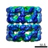
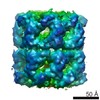
Journal: Structure / Year: 2008 Title: De novo backbone trace of GroEL from single particle electron cryomicroscopy. Authors: Steven J Ludtke / Matthew L Baker / Dong-Hua Chen / Jiu-Li Song / David T Chuang / Wah Chiu / Abstract: In this work, we employ single-particle electron cryo-microscopy (cryo-EM) to reconstruct GroEL to approximately 4 A resolution with both D7 and C7 symmetry. Using a newly developed skeletonization ...In this work, we employ single-particle electron cryo-microscopy (cryo-EM) to reconstruct GroEL to approximately 4 A resolution with both D7 and C7 symmetry. Using a newly developed skeletonization algorithm and secondary structure element identification in combination with sequence-based secondary structure prediction, we demonstrate that it is possible to achieve a de novo Calpha trace directly from a cryo-EM reconstruction. The topology of our backbone trace is completely accurate, though subtle alterations illustrate significant differences from existing crystal structures. In the map with C7 symmetry, the seven monomers in each ring are identical; however, the subunits have a subtly different structure in each ring, particularly in the equatorial domain. These differences include an asymmetric salt bridge, density in the nucleotide-binding pocket of only one ring, and small shifts in alpha helix positions. This asymmetric conformation is different from previous asymmetric structures, including GroES-bound GroEL, and may represent a "primed state" in the chaperonin pathway.
History
Deposition
Jan 25, 2008
-
Header (metadata) release
Feb 12, 2008
-
Map release
Apr 14, 2009
-
Update
Nov 9, 2016
-
Current status
Nov 9, 2016
Processing site: RCSB / Status: Released
-
Structure visualization
Movie
Surface view with section colored by density value
GO: protein folding / InterPro: INTERPRO: IPR012723
-
Experimental details
-
Structure determination
Method
cryo EM
Processing
single particle reconstruction
Aggregation state
particle
-
Sample preparation
Buffer
Details: 20 mM Tris.HCl, pH 7.5, 50 mM MgCl2
Grid
Details: Quantifoil grids with 2 um holes
Vitrification
Cryogen name: ETHANE / Chamber humidity: 100 % / Chamber temperature: 100 K / Instrument: OTHER / Details: Vitrification instrument: Vitrobot / Method: Blot for 2 sec
-
Electron microscopy
Microscope
JEOL 3000SFF
Temperature
Min: 4 K / Max: 4 K / Average: 4 K
Details
low dose on JEOL 3000SFF
Date
Jan 1, 2005
Image recording
Category: FILM / Film or detector model: KODAK SO-163 FILM / Digitization - Scanner: NIKON SUPER COOLSCAN 9000 / Digitization - Sampling interval: 6.35 µm / Number real images: 135 / Average electron dose: 36 e/Å2 / Bits/pixel: 14
Electron beam
Acceleration voltage: 300 kV / Electron source: FIELD EMISSION GUN
In the structure databanks used in Yorodumi, some data are registered as the other names, "COVID-19 virus" and "2019-nCoV". Here are the details of the virus and the list of structure data.
Jan 31, 2019. EMDB accession codes are about to change! (news from PDBe EMDB page)
EMDB accession codes are about to change! (news from PDBe EMDB page)
The allocation of 4 digits for EMDB accession codes will soon come to an end. Whilst these codes will remain in use, new EMDB accession codes will include an additional digit and will expand incrementally as the available range of codes is exhausted. The current 4-digit format prefixed with “EMD-” (i.e. EMD-XXXX) will advance to a 5-digit format (i.e. EMD-XXXXX), and so on. It is currently estimated that the 4-digit codes will be depleted around Spring 2019, at which point the 5-digit format will come into force.
The EM Navigator/Yorodumi systems omit the EMD- prefix.
Related info.:Q: What is EMD? / ID/Accession-code notation in Yorodumi/EM Navigator
Yorodumi is a browser for structure data from EMDB, PDB, SASBDB, etc.
This page is also the successor to EM Navigator detail page, and also detail information page/front-end page for Omokage search.
The word "yorodu" (or yorozu) is an old Japanese word meaning "ten thousand". "mi" (miru) is to see.
Related info.:EMDB / PDB / SASBDB / Comparison of 3 databanks / Yorodumi Search / Aug 31, 2016. New EM Navigator & Yorodumi / Yorodumi Papers / Jmol/JSmol / Function and homology information / Changes in new EM Navigator and Yorodumi
 Movie
Movie Controller

 Yorodumi
 Open data
 Basic information
 Map data
 Sample
 Keywords
 Function and homology information
 Authors
 Citation
 Structure visualization
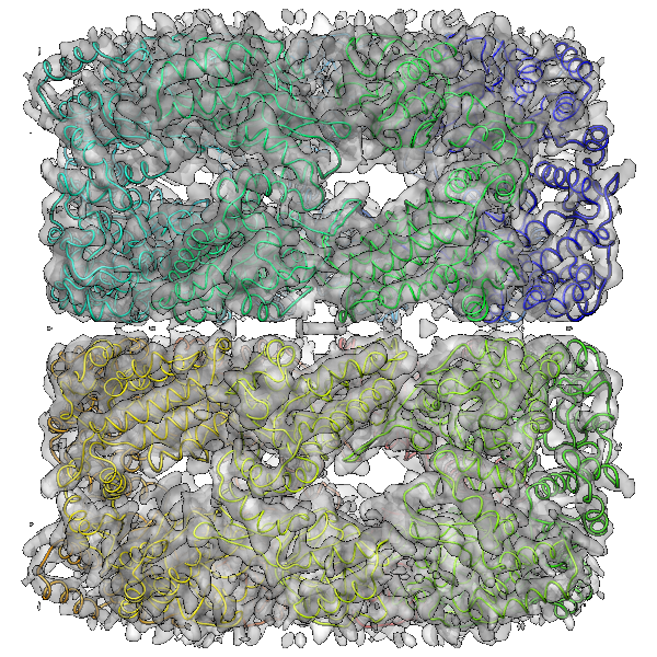
 Downloads & links
 emd_5001_1.png
 http://ftp.pdbj.org/pub/emdb/structures/EMD-5001


 Z (Sec.)
 Y (Row.)
 X (Col.)


 Sample components
 Processing
 Electron microscopy
 FIELD EMISSION GUN