 ムービー
ムービー コントローラー


+ データを開く

- 基本情報
基本情報
| 登録情報 | データベース: EMDB / ID: EMD-5001 | |||||||||
|---|---|---|---|---|---|---|---|---|---|---|
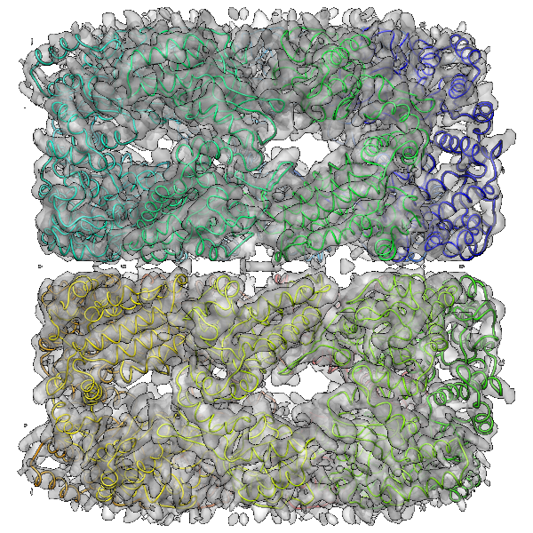
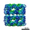
| タイトル | Native, unliganded GroEL, D7 symmetrized, 4.2 A resolution 0.5 criterion | |||||||||
 マップデータ マップデータ | D7 structure of GroEL at 4.2 Angstrom resolution | |||||||||
 試料 試料 |
| |||||||||
 キーワード キーワード |  groel / chaperonin / chaperone (シャペロン) / backbone trace / eman / single particle (単粒子解析法) groel / chaperonin / chaperone (シャペロン) / backbone trace / eman / single particle (単粒子解析法) | |||||||||
| 機能・相同性 |  機能・相同性情報 機能・相同性情報GroEL-GroES complex / chaperonin ATPase / virion assembly / chaperone cofactor-dependent protein refolding / isomerase activity / ATP-dependent protein folding chaperone / response to radiation / unfolded protein binding / フォールディング / response to heat ...GroEL-GroES complex / chaperonin ATPase / virion assembly / chaperone cofactor-dependent protein refolding / isomerase activity / ATP-dependent protein folding chaperone / response to radiation / unfolded protein binding / フォールディング / response to heat / protein refolding / magnesium ion binding / ATP hydrolysis activity / ATP binding / 生体膜 / identical protein binding / 細胞質基質類似検索 - 分子機能 | |||||||||
| 生物種 |  Escherichia coli (大腸菌) Escherichia coli (大腸菌) | |||||||||
| 手法 | 単粒子再構成法 / クライオ電子顕微鏡法 / 解像度: 4.2 Å | |||||||||
 データ登録者 データ登録者 | Ludtke SJ / Baker ML / Chen D / Song J / Chuang DT / Chiu W | |||||||||
 引用 引用 | ジャーナル: Structure / 年: 2008 タイトル: De novo backbone trace of GroEL from single particle electron cryomicroscopy. 著者: Steven J Ludtke / Matthew L Baker / Dong-Hua Chen / Jiu-Li Song / David T Chuang / Wah Chiu /  要旨: In this work, we employ single-particle electron cryo-microscopy (cryo-EM) to reconstruct GroEL to approximately 4 A resolution with both D7 and C7 symmetry. Using a newly developed skeletonization ...In this work, we employ single-particle electron cryo-microscopy (cryo-EM) to reconstruct GroEL to approximately 4 A resolution with both D7 and C7 symmetry. Using a newly developed skeletonization algorithm and secondary structure element identification in combination with sequence-based secondary structure prediction, we demonstrate that it is possible to achieve a de novo Calpha trace directly from a cryo-EM reconstruction. The topology of our backbone trace is completely accurate, though subtle alterations illustrate significant differences from existing crystal structures. In the map with C7 symmetry, the seven monomers in each ring are identical; however, the subunits have a subtly different structure in each ring, particularly in the equatorial domain. These differences include an asymmetric salt bridge, density in the nucleotide-binding pocket of only one ring, and small shifts in alpha helix positions. This asymmetric conformation is different from previous asymmetric structures, including GroES-bound GroEL, and may represent a "primed state" in the chaperonin pathway. | |||||||||
| 履歴 |
|
- 構造の表示
構造の表示
| ムービー |
ムービービューア |
|---|---|
| 構造ビューア | EMマップ: SurfViewMolmilJmol/JSmol |
| 添付画像 |
- ダウンロードとリンク
-EMDBアーカイブ
| マップデータ | emd_5001.map.gz | 13.7 MB | EMDBマップデータ形式 | |
|---|---|---|---|---|
| ヘッダ (付随情報) | emd-5001-v30.xmlemd-5001.xml | 10 KB 10 KB | 表示 表示 | EMDBヘッダ |
| 画像 |  emd_5001_1.png emd_5001_1.png | 628.1 KB | ||
| アーカイブディレクトリ |  http://ftp.pdbj.org/pub/emdb/structures/EMD-5001ftp://ftp.pdbj.org/pub/emdb/structures/EMD-5001 http://ftp.pdbj.org/pub/emdb/structures/EMD-5001ftp://ftp.pdbj.org/pub/emdb/structures/EMD-5001 | HTTPS FTP |
-関連構造データ

-リンク
| EMDBのページ | EMDB (EBI/PDBe) / EMDataResource |
|---|---|
| 「今月の分子」の関連する項目 |

-マップ
| ファイル | ダウンロード / ファイル: emd_5001.map.gz / 形式: CCP4 / 大きさ: 29.8 MB / タイプ: IMAGE STORED AS FLOATING POINT NUMBER (4 BYTES) | ||||||||||||||||||||||||||||||||||||||||||||||||||||||||||||
|---|---|---|---|---|---|---|---|---|---|---|---|---|---|---|---|---|---|---|---|---|---|---|---|---|---|---|---|---|---|---|---|---|---|---|---|---|---|---|---|---|---|---|---|---|---|---|---|---|---|---|---|---|---|---|---|---|---|---|---|---|---|
| 注釈 | D7 structure of GroEL at 4.2 Angstrom resolution | ||||||||||||||||||||||||||||||||||||||||||||||||||||||||||||
| ボクセルのサイズ | X=Y=Z: 1.06 Å | ||||||||||||||||||||||||||||||||||||||||||||||||||||||||||||
| 密度 |
| ||||||||||||||||||||||||||||||||||||||||||||||||||||||||||||
| 対称性 | 空間群: 1 | ||||||||||||||||||||||||||||||||||||||||||||||||||||||||||||
| 詳細 | EMDB XML:
CCP4マップ ヘッダ情報:
| ||||||||||||||||||||||||||||||||||||||||||||||||||||||||||||
-添付データ
- 試料の構成要素
-全体 : Native unliganded GroEL, residual ADP
| 全体 | 名称: Native unliganded GroEL, residual ADP |
|---|---|
| 要素 |
|
-超分子 #1000: Native unliganded GroEL, residual ADP
| 超分子 | 名称: Native unliganded GroEL, residual ADP / タイプ: sample / ID: 1000 / 集合状態: Two back to back homo-heptameric rings / Number unique components: 1 |
|---|---|
| 分子量 | 理論値: 800 KDa |
-分子 #1: GroEL
| 分子 | 名称: GroEL / タイプ: protein_or_peptide / ID: 1 / Name.synonym: GroEL / コピー数: 14 / 集合状態: 14-mer / 組換発現: Yes |
|---|---|
| 由来(天然) | 生物種: Escherichia coli (大腸菌) |
| 分子量 | 理論値: 800 KDa |
| 組換発現 | 生物種: Escherichia coli (大腸菌) / 組換株: ESts CG-712 / 組換プラスミド: pGroESL |
| 配列 | GO: フォールディング / InterPro: INTERPRO: IPR012723 |
-実験情報
-構造解析
| 手法 | クライオ電子顕微鏡法 |
|---|---|
 解析 解析 | 単粒子再構成法 |
| 試料の集合状態 | particle |
-試料調製
| 緩衝液 | 詳細: 20 mM Tris.HCl, pH 7.5, 50 mM MgCl2 |
|---|---|
| グリッド | 詳細: Quantifoil grids with 2 um holes |
| 凍結 | 凍結剤: ETHANE / チャンバー内湿度: 100 % / チャンバー内温度: 100 K / 装置: OTHER / 詳細: Vitrification instrument: Vitrobot / 手法: Blot for 2 sec |
- 電子顕微鏡法
| 顕微鏡 | JEOL 3000SFF |
|---|---|
| 電子線 | 加速電圧: 300 kV / 電子線源: FIELD EMISSION GUN |
| 電子光学系 | 照射モード: FLOOD BEAM / 撮影モード: OTHER / Cs: 1.6 mm / 最大 デフォーカス(公称値): 2.3 µm / 最小 デフォーカス(公称値): 0.9 µm / 倍率(公称値): 60000 |
| 試料ステージ | 試料ホルダー: Top entry / 試料ホルダーモデル: OTHER |
| 温度 | 最低: 4 K / 最高: 4 K / 平均: 4 K |
| 詳細 | low dose on JEOL 3000SFF |
| 日付 | 2005年1月1日 |
| 撮影 | カテゴリ: FILM / フィルム・検出器のモデル: KODAK SO-163 FILM デジタル化 - スキャナー: NIKON SUPER COOLSCAN 9000 デジタル化 - サンプリング間隔: 6.35 µm / 実像数: 135 / 平均電子線量: 36 e/Å2 / ビット/ピクセル: 14 |
-画像解析
| CTF補正 | 詳細: per micrograph |
|---|---|
| 最終 再構成 | アルゴリズム: OTHER / 解像度のタイプ: BY AUTHOR / 解像度: 4.2 Å / 解像度の算出法: FSC 0.5 CUT-OFF / ソフトウェア - 名称: EMAN / 使用した粒子像数: 20401 |
