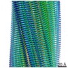
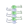
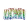
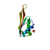
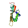
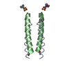
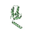
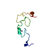
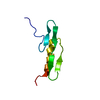
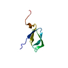
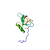
ジャーナル: Nat Struct Mol Biol / 年: 2018 タイトル: Atomic-level evidence for packing and positional amyloid polymorphism by segment from TDP-43 RRM2. 著者: Elizabeth L Guenther / Peng Ge / Hamilton Trinh / Michael R Sawaya / Duilio Cascio / David R Boyer / Tamir Gonen / Z Hong Zhou / David S Eisenberg / 要旨: Proteins in the fibrous amyloid state are a major hallmark of neurodegenerative disease. Understanding the multiple conformations, or polymorphs, of amyloid proteins at the molecular level is a ...Proteins in the fibrous amyloid state are a major hallmark of neurodegenerative disease. Understanding the multiple conformations, or polymorphs, of amyloid proteins at the molecular level is a challenge of amyloid research. Here, we detail the wide range of polymorphs formed by a segment of human TAR DNA-binding protein 43 (TDP-43) as a model for the polymorphic capabilities of pathological amyloid aggregation. Using X-ray diffraction, microelectron diffraction (MicroED) and single-particle cryo-EM, we show that the DLIIKGISVHI segment from the second RNA-recognition motif (RRM2) forms an array of amyloid polymorphs. These associations include seven distinct interfaces displaying five different symmetry classes of steric zippers. Additionally, we find that this segment can adopt three different backbone conformations that contribute to its polymorphic capabilities. The polymorphic nature of this segment illustrates at the molecular level how amyloid proteins can form diverse fibril structures.
 ムービー
ムービー コントローラー


 データを開く
 基本情報
基本情報 要素
要素 TAR DNA結合タンパク質43
  キーワード
キーワード 機能・相同性情報
 データ登録者
 米国, 1件
  引用
引用 構造の表示
構造の表示 ダウンロードとリンク
 その他のダウンロード
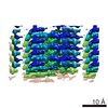
 PDBj
PDBj
 集合体
集合体
 試料調製
試料調製 解析
解析