 Movie
Movie Controller

[English] 日本語
 Yorodumi
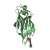
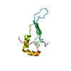
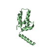
- PDB-5uvr: The core region of PilO from the type IV pilus system of Pseudomo... -
+ Open data

- Basic information
| Entry | Database: PDB / ID: 5uvr | ||||||
|---|---|---|---|---|---|---|---|
| Title | The core region of PilO from the type IV pilus system of Pseudomonas aeruginosa | ||||||
 Components Components | PilO protein | ||||||
 Keywords Keywords | MEMBRANE PROTEIN / alignment subcomplex / modified ferredoxin fold / reductive methylation / type iv pili | ||||||
| Function / homology |  Function and homology information Function and homology informationoligosaccharyl transferase activity / type IV pilus assembly / type IV pilus-dependent motility Similarity search - Function | ||||||
| Biological species |   Pseudomonas aeruginosa (bacteria) Pseudomonas aeruginosa (bacteria) | ||||||
| Method |  X-RAY DIFFRACTION / SYNCHROTRON / MOLECULAR REPLACEMENT / Resolution: 1.7 Å X-RAY DIFFRACTION / SYNCHROTRON / MOLECULAR REPLACEMENT / Resolution: 1.7 Å | ||||||
 Authors Authors | Howell, P.L. / Junop, M.S. | ||||||
| Funding support |  Canada, 1items Canada, 1items
| ||||||
 Citation Citation | Journal: Sci Rep / Year: 2018 Title: Conserved, unstructured regions in Pseudomonas aeruginosa PilO are important for type IVa pilus function. Authors: Leighton, T.L. / Mok, M.C. / Junop, M.S. / Howell, P.L. / Burrows, L.L. | ||||||
| History |
|
- Structure visualization
| Structure viewer | Molecule: MolmilJmol/JSmol |
|---|
- Downloads & links
-Download
| PDBx/mmCIF format | 5uvr.cif.gz | 35.6 KB | Display | PDBx/mmCIF format |
|---|---|---|---|---|
| PDB format | pdb5uvr.ent.gz | 22.4 KB | Display | PDB format |
| PDBx/mmJSON format | 5uvr.json.gz | Tree view | PDBx/mmJSON format | |
| Others |  Other downloads Other downloads |
-Validation report
| Arichive directory | https://data.pdbj.org/pub/pdb/validation_reports/uv/5uvrftp://data.pdbj.org/pub/pdb/validation_reports/uv/5uvr | HTTPS FTP |
|---|
-Related structure data
| Related structure data |  2rjzS S: Starting model for refinement |
|---|---|
| Similar structure data |


-Links
 PDBj
PDBj
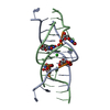
- Assembly
| Deposited unit | 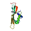
| ||||||||||||
|---|---|---|---|---|---|---|---|---|---|---|---|---|---|
| 1 |
| ||||||||||||
| 2 | 
| ||||||||||||
| Unit cell |
| ||||||||||||
| Components on special symmetry positions |
|
-Components
| #1: Protein | Mass: 10813.502 Da / Num. of mol.: 1 / Fragment: core region (UNP residues 109-206) Source method: isolated from a genetically manipulated source Source: (gene. exp.) Pseudomonas aeruginosa (bacteria)Gene: pilO, PAERUG_E15_London_28_01_14_03389, PAERUG_P32_London_17_VIM_2_10_11_01500 Production host: |
|---|---|
| #2: Water | ChemComp-HOH /  Mass: 18.015 Da / Num. of mol.: 97 / Source method: isolated from a natural source / Formula: H2O Mass: 18.015 Da / Num. of mol.: 97 / Source method: isolated from a natural source / Formula: H2O |
-Experimental details
-Experiment
| Experiment | Method: X-RAY DIFFRACTION / Number of used crystals: 1 |
|---|
- Sample preparation
| Crystal | Density Matthews: 2.78 Å3/Da / Density % sol: 55.82 % |
|---|---|
| Crystal grow | Temperature: 277 K / Method: vapor diffusion, hanging drop Details: 0.2 M NaCl, 0.1 M CAPS pH 10.5, 20% (v/v) PEG 8000), 3% (v/v) DMSO |
-Data collection
| Diffraction | Mean temperature: 100 K |
|---|---|
| Diffraction source | Source: SYNCHROTRON / Site: CLSI / Beamline: 08ID-1 / Wavelength: 0.979 Å |
| Detector | Type: MARMOSAIC 300 mm CCD / Detector: CCD / Date: Oct 25, 2014 |
| Radiation | Protocol: SINGLE WAVELENGTH / Monochromatic (M) / Laue (L): M / Scattering type: x-ray |
| Radiation wavelength | Wavelength: 0.979 Å / Relative weight: 1 |
| Reflection | Resolution: 1.7→35.342 Å / Num. obs: 14759 / % possible obs: 99.9 % / Redundancy: 6.7 % / Net I/σ(I): 10.9 |
- Processing
| Software |
| |||||||||||||||||||||||||||||||||||||||||||||||||||||||||||||||||||||||||||||
|---|---|---|---|---|---|---|---|---|---|---|---|---|---|---|---|---|---|---|---|---|---|---|---|---|---|---|---|---|---|---|---|---|---|---|---|---|---|---|---|---|---|---|---|---|---|---|---|---|---|---|---|---|---|---|---|---|---|---|---|---|---|---|---|---|---|---|---|---|---|---|---|---|---|---|---|---|---|---|
| Refinement | Method to determine structure: MOLECULAR REPLACEMENT Starting model: 2RJZ Resolution: 1.7→35.342 Å / SU ML: 0.15 / Cross valid method: FREE R-VALUE / σ(F): 1.35 / Phase error: 26.64
| |||||||||||||||||||||||||||||||||||||||||||||||||||||||||||||||||||||||||||||
| Solvent computation | Shrinkage radii: 0.9 Å / VDW probe radii: 1.11 Å | |||||||||||||||||||||||||||||||||||||||||||||||||||||||||||||||||||||||||||||
| Refinement step | Cycle: LAST / Resolution: 1.7→35.342 Å
| |||||||||||||||||||||||||||||||||||||||||||||||||||||||||||||||||||||||||||||
| Refine LS restraints |
| |||||||||||||||||||||||||||||||||||||||||||||||||||||||||||||||||||||||||||||
| LS refinement shell |
|
